ELISA Kits
Showing 1051–1200 of 3623 results
-

HLA-DR (α and β chains) ELISA Standard has been tested and formulated to work exclusively with Cayman’s HLA-DR (α and β chains) ELISA Kit (Item No. 501810). Please visit HLA-DR (α and β chains) ELISA Kit (Item No. 501810) for the kit protocol, procedures, and product handling.
Brand:CaymanSKU:480131 - 200 ngAvailable on backorder
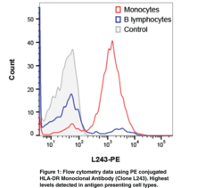
HLA-DR is a MHC Class II cell surface receptor heterodimer composed of a 33-35 kDa α chain, a ~30 kDa β chain, and a 10-30 amino acid ligand.{33426} When the heterodimer is fully combined on the cell surface of an antigen-presenting cell, such as macrophages, B cells, and dendritic cells, they present that ligand primarily to CD-4+ T cells.{15687} This presentation coupled with the T cell response can stimulate or suppress an antibody response to that ligand.{15687} HLR-DRs have been linked to a number of autoimmune disorders such as rheumatoid arthritis, lupus, and psoriasis as well as diabetes, hepatitis, and sclerosis among a number of others.{34485,34486,34487,34488,34489,34490,34491} Cayman’s HLA-DR Monoclonal Antibody (Clone L243) detects HLA-DR with or without attached ligand. The predicated size of the HLA-DRα or HLA-DRβ subunits is ~29-30 kDa.
Brand:CaymanSKU:21827 - 100 µgAvailable on backorder
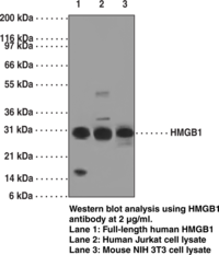
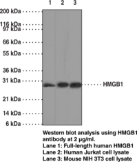
High Mobility Group Protein B1 (HMGB1) belongs to the HMGB family and contains two HMG box DNA-binding domains. It is a highly conserved, ubiquitous protein present in the nuclei and cytoplasm of nearly all cell types, and is a necessary and sufficient mediator of inflammation during sterile and infection-associated responses. HMGB1 also act as DNA nuclear binding protein that has recently been shown to be an early trigger of sterile inflammation in animal models of trauma-hemorrhage via the activation of the Toll-like receptor 4 (TLR4) and the receptor for the advanced glycation endproducts (RAGE).{20300} It is reported that the level of HMGB1 is elevated during sterile tissue injury, infection, lethal endotoxemia or sepsis, collagen-induced arthritis, and ischemia-reperfusion induced tissue injury.{20304}
Brand:CaymanSKU:11512 - 1 eaAvailable on backorder
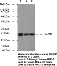
High Mobility Group Protein B1 (HMGB1) belongs to the HMGB family and contains two HMG box DNA-binding domains. It is a highly conserved, ubiquitous protein present in the nuclei and cytoplasm of nearly all cell types, and is a necessary and sufficient mediator of inflammation during sterile and infection-associated responses. HMGB1 also act as DNA nuclear binding protein that has been shown to be an early trigger of sterile inflammation in animal models of trauma-hemorrhage via the activation of the Toll-like receptor 4 (TLR4) and the receptor for the advanced glycation endproducts (RAGE).{20300} It is reported that the level of HMGB1 is elevated during sterile tissue injury, infection, lethal endotoxemia or sepsis, collagen-induced arthritis, and ischemia-reperfusion induced tissue injury.{20304}
Brand:CaymanSKU:11514 - 1 eaAvailable on backorder
High Mobility Group Protein B1 (HMGB1) belongs to the HMGB family and contains two HMG box DNA-binding domains. It is a highly conserved, ubiquitous protein present in the nuclei and cytoplasm of nearly all cell types, and is a necessary and sufficient mediator of inflammation during sterile and infection-associated responses. HMGB1 also act as DNA nuclear binding protein that has been shown to be an early trigger of sterile inflammation in animal models of trauma-hemorrhage via the activation of the Toll-like receptor 4 (TLR4) and the receptor for the advanced glycation endproducts (RAGE).{20300} It is reported that the level of HMGB1 is elevated during sterile tissue injury, infection, lethal endotoxemia or sepsis, collagen-induced arthritis, and ischemia-reperfusion induced tissue injury.{20304}
Brand:CaymanSKU:11513 - 1 eaAvailable on backorder

High Mobility Group Protein B1 (HMGB1) belongs to the HMGB family and contains two HMG box DNA-binding domains. It is a highly conserved, ubiquitous protein present in the nuclei and cytoplasm of nearly all cell types, and is a necessary and sufficient mediator of inflammation during sterile and infection-associated responses. HMGB1 also act as DNA nuclear binding protein that has recently been shown to be an early trigger of sterile inflammation in animal models of trauma-hemorrhage via the activation of the Toll-like receptor 4 (TLR4) and the receptor for the advanced glycation endproducts (RAGE).{20300} It is reported that the level of HMGB1 is elevated during sterile tissue injury, infection, lethal endotoxemia or sepsis, collagen-induced arthritis, and ischemia-reperfusion induced tissue injury.{20304}
Brand:CaymanSKU:11515 - 1 eaAvailable on backorder
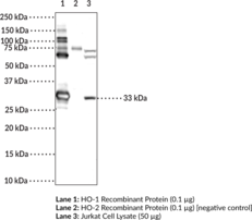
Heme oxygenase-1 (HO-1), also known as heat shock protein 32 (Hsp32), is an inducible heme oxygenase encoded by the HMOX1 gene.{40053,40052,38123} It is a membrane-bound enzyme that catalyzes the cleavage of heme to release carbon monoxide (CO), ferrous ions (Fe2+), and biliverdin, with biliverdin being further processed into bilirubin. HO-1 is found in human spleen, liver, and kidney where its expression is induced by the presence of heme, hormones, metals, oxidative agents, and therapeutic compounds to protect against oxidative stress and inflammatory responses. HO-1 is upregulated in a variety of cancers and siRNA knockdown of HMOX1 or inhibition of HO-1 decreases cancer cell proliferation.{38124,38125,38126} HO-1 also interacts with the severe acute respiratory syndrome coronavirus 2 (SARS-CoV-2) accessory protein Orf3a that, in a similar virus, SARS-CoV, is associated with activation of the NLRP3 inflammasome.{55024,55036,55037} Cayman’s HO-1 (Hsp32) Polyclonal Antibody can be used for ELISA, immunofluorescence (IF), immunohistochemistry (IHC), and Western blot (WB) applications. The antibody recognizes HO-1 (Hsp32) at approximately 32 kDa from human samples.
Brand:CaymanSKU:24633 - 500 µgAvailable on backorder
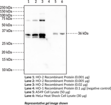
Heme oxygenase-2 (HO-2) is a constitutively active heme oxygenase encoded by the HMOX2 gene.{6104,40054} It is a membrane-bound enzyme that catalyzes the cleavage of heme to give carbon monoxide (CO), ferrous ions (Fe2+), and biliverdin, with biliverdin being further processed into bilirubin. HO-2 is found in neurons, testes, and endothelial and smooth muscle cells from cerebral vessels.{40054} HO-2 protects against apoptotic neuronal cell death in models of ischemic injury and oxidative stress. It also acts as an oxygen sensor to inhibit systemic vascular reactivity and reduce cell death in response to hypoxia. Cayman’s HO-2 Monoclonal Antibody (Clone 5D10) can be used for Western blot and ELISA applications. The antibody recognizes HO-2 at 36 kDa from human samples.
Brand:CaymanSKU:25691 - 500 µgAvailable on backorder
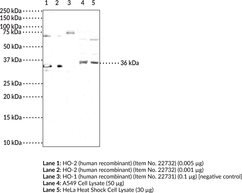
Heme oxygenase-2 (HO-2) is a constitutively active heme oxygenase encoded by the HMOX2 gene.{6104,40054} It is a membrane-bound enzyme that catalyzes the cleavage of heme to give carbon monoxide (CO), ferrous ions (Fe2+), and biliverdin, with biliverdin being further processed into bilirubin. HO-2 is found in neurons, testes, and endothelial and smooth muscle cells from cerebral vessels.{40054} HO-2 protects against apoptotic neuronal cell death in models of ischemic injury and oxidative stress. It also acts as an oxygen sensor to inhibit systemic vascular reactivity and reduce cell death in response to hypoxia. Cayman’s HO-2 Polyclonal Antibody can be used for Western blot and ELISA applications. The antibody recognizes HO-2 at 36 kDa from human samples.
Brand:CaymanSKU:24634 - 500 µgAvailable on backorder
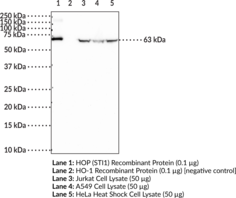
Hsp70/Hsp90 organizing protein (HOP), also known as stress-induced phosphoprotein 1 (STI1 or STIP1), is a co-chaperone that reversibly forms a complex with heat shock protein 70 (Hsp70) and Hsp90 during the Hsp90 chaperone cycle to facilitate the transfer of client proteins from Hsp70 to Hsp90. It contains three tetratricopeptide repeat domains (TPR1, TPR2A, and TPR2B) that act as binding regions, with TPR1 and TPR2B binding to Hsp70 (Item Nos. 22739 | 23002) and TPR2A binding to Hsp90 (Item Nos. 22734 | 22735).{39121} HOP is both a nuclear and cytoplasmic protein, capable of travel between both.{39122} HOP has also been found to interact with several other proteins and complexes, including but not limited to Hsc70 (Item No. 22737), PACRG, METTL21B, FLCN, FNIP1, and FNIP2.{39123,39124,39125,39126,39127} Cayman’s HOP (STI1) Polyclonal Antibody detects HOP (STI1) at 63 kDa.
Brand:CaymanSKU:24529 - 100 µgAvailable on backorder

Hormone-sensitive lipase (HSL) catalyzes the hydrolysis of tri-, di-, and monoacylglycerols, as well as cholesterol esters and thus mobilizes fatty acid and provides a primary source of energy in mammals.{13168} The enzyme is highly expressed in adipose tissue and steroidogenic tissues, and less abundantly in skeletal muscle, heart, brain, pancreatic beta cells, adrenal gland, ovaries, testes, and macrophages. Its presence in various tissues indicates the enzyme plays diverse roles including those in steroidogenesis and spermatogenesis, foam cell formation in atherosclerosis, and diabetic pathology.{13168,13167} Human HSL cDNA encodes a 775 amino acid protein with an estimated molecular size of 84 kDa.{13169} A second, larger isoform encoded by a unique testis mRNA was later identified.{13171,13166} Cayman’s HSL Polyclonal Antibody can be used for Western blot applications. The antibody recognizes HSL at 86 kDa from human, mouse, and rat samples.
Brand:CaymanSKU:10006371 - 500 µlAvailable on backorder
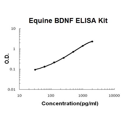
Sandwich High Sensitivity ELISA kit for Quantitative Detection of horse equine BDNF. 96wells/kit, with removable strips.
Brand:Boster BioSKU:EK0307-EQAvailable on backorder
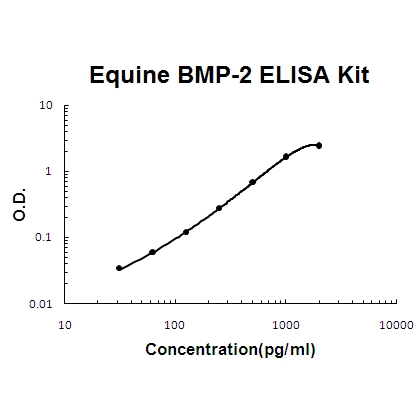
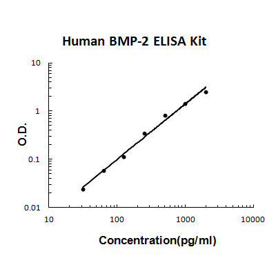
Sandwich High Sensitivity ELISA kit for Quantitative Detection of horse equine BMP-2. 96wells/kit, with removable strips.
Brand:Boster BioSKU:EK0311-EQAvailable on backorder
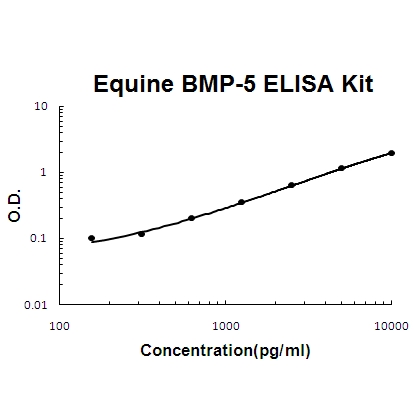
Sandwich High Sensitivity ELISA kit for Quantitative Detection of horse equine BMP-5. 96wells/kit, with removable strips.
Brand:Boster BioSKU:EK0310-EQAvailable on backorder
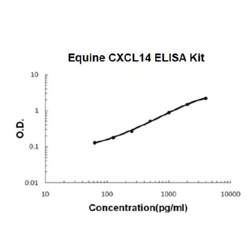
Sandwich High Sensitivity ELISA kit for Quantitative Detection of horse equine CXCL14. 96wells/kit, with removable strips.
Brand:Boster BioSKU:EK1285-EQAvailable on backorder
Sandwich High Sensitivity ELISA kit for Quantitative Detection of horse equine Endothelin. 96wells/kit, with removable strips.
Brand:Boster BioSKU:EK0945-EQAvailable on backorder
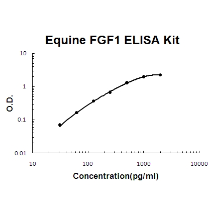
Sandwich High Sensitivity ELISA kit for Quantitative Detection of horse equine FGF1. 96wells/kit, with removable strips.
Brand:Boster BioSKU:EK0339-EQAvailable on backorder
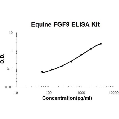
Sandwich High Sensitivity ELISA kit for Quantitative Detection of horse equine FGF9. 96wells/kit, with removable strips.
Brand:Boster BioSKU:EK0348-EQAvailable on backorder
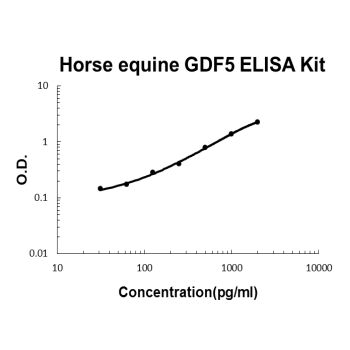
Sandwich High Sensitivity ELISA kit for Quantitative Detection of horse equine GDF5. 96wells/kit, with removable strips.
Brand:Boster BioSKU:EK1504-EQAvailable on backorder
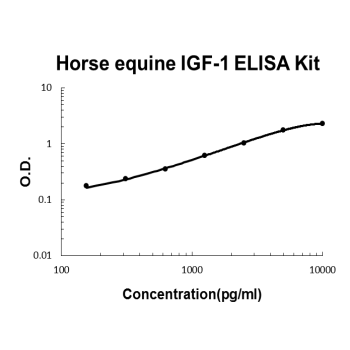
Sandwich High Sensitivity ELISA kit for Quantitative Detection of horse equine IGF-1. 96wells/kit, with removable strips.
Brand:Boster BioSKU:EK0376-EQAvailable on backorder
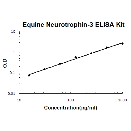
Sandwich High Sensitivity ELISA kit for Quantitative Detection of horse equine Neurotrophin-3. 96wells/kit, with removable strips.
Brand:Boster BioSKU:EK0472-EQAvailable on backorder
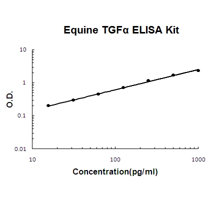
Sandwich High Sensitivity ELISA kit for Quantitative Detection of horse equine TGF alpha. 96wells/kit, with removable strips.
Brand:Boster BioSKU:EK0511-EQAvailable on backorder
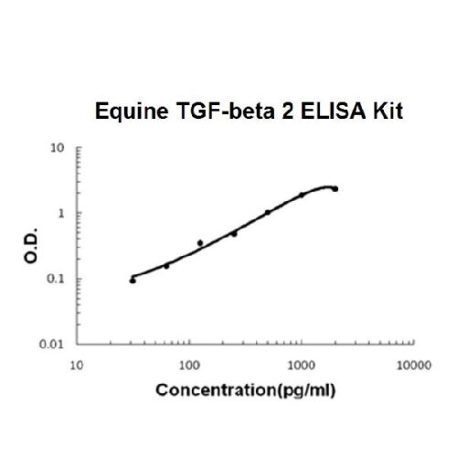
Sandwich High Sensitivity ELISA kit for Quantitative Detection of activated horse equine TGF-beta 2. 96wells/kit, with removable strips.
Brand:Boster BioSKU:EK0981-EQAvailable on backorder
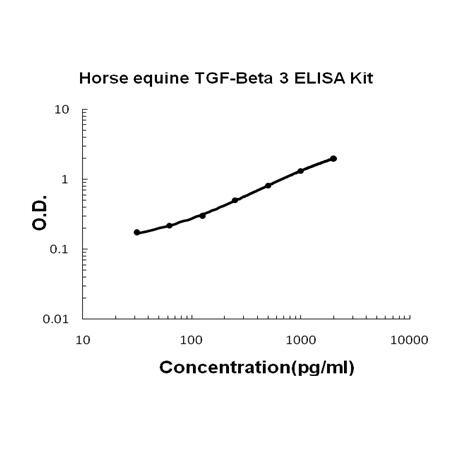
Sandwich High Sensitivity ELISA kit for Quantitative Detection of activated horse equine TGF-beta 3. 96wells/kit, with removable strips.
Brand:Boster BioSKU:EK1103-EQAvailable on backorder
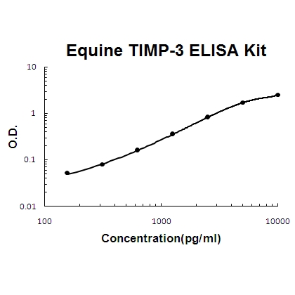
Sandwich High Sensitivity ELISA kit for Quantitative Detection of horse equine TIMP-3. 96wells/kit, with removable strips.
Brand:Boster BioSKU:EK0523-EQAvailable on backorder
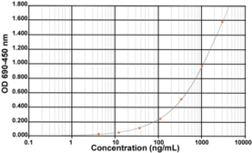
Impurity assessment is a key step during the drug development production of recombinant proteins, including therapeutic proteins. Specific impurities coming from the cells mediating the protein expression, known as ‘Host Cell Proteins (HCP)’, are generated and need to be removed. This kit is intended for use in assessing relative quantities of E. coli HCP in manufactured or research bioproducts. Polyclonal antibodies used in this kit have been generated against several strains of E. coli and specifically selected for their recognition of a large spectrum of E. coli proteins. Thus, this kit can be considered as generic and allows a relative-quantitative determination of E. coli HCP in many types of samples, such as samples issued from the purification process (HCP clearance), process control, quality control, or product release. Using this kit, HCP concentration is measured in ng/ml (HCP equivalent is extrapolated from a standard curve). Conventionally, the HCP content in a product will finally be expressed in ng/mg, where ng represents HCP mass and mg represents the product mass. Note that contrary to the concentration measurement of the product, the HCP signal is only reflective of antibody binding and does not strictly reflect the mass of HCP. This kit has been successfully validated for recovery and precision using reconstituted HCP samples and tested against different final products. Given the diversity of final products, all potential matrix effects cannot be known and it is recommended that you test the suitability of the kit for use with your own HCP samples in your laboratory. This kit should be used as one part of your complete HCP analysis. [Bertin Catalog No. A05034]
Brand:CaymanSKU:18919 - 96 wellsAvailable on backorder
Brand:CaymanSKU:660910 - 15 mlAvailable on backorder
Heat shock factor 1 (Hsf1) belongs to a family of heat shock transcription factors that activate the transcription of genes encoding products required for protein folding, processing, targeting, degradation, and function.{15655} The up-regulation of Hsp expression by stressors is achieved at the level of transcription through a heat shock element (HSE) and a transcription factor.{15642,15643,15644} Most Hsfs have highly conserved amino acid sequences. On all Hsfs there is a DNA binding domain at the N-terminus. Hydrophobic repeats located adjacent to this binding domain are essential for the formation of active trimers. Towards the C-terminal region another short hydrophobic repeat exists and is thought to be necessary for suppression of trimerization.{15645} There are two main Hsfs, 1 and 2. Murine Hsf1 exists as two isoforms, however in higher eukaryotes Hsf1 is found in a diffuse cytoplasmic and nuclear distribution in unstressed cells. Once exposed to a multitude of stressors, it localizes to discrete nuclear granules within seconds. As it recovers from stress, Hsf1 dissipates from these granules to a diffuse nucleoplasmic distribution. Hsf2 on the other hand is similar to murine Hsf1, as it exists as two isoforms, the α form being more transcriptionally active than the smaller β form.{15656,15647} Various experiments have suggested that Hsf2 may have roles in differentiation and development.{15648,15649,15650}
Brand:CaymanSKU:10011433 - 100 µgAvailable on backorder
Heat shock factor 1 (Hsf1) belongs to a family of heat shock transcription factors that activate the transcription of genes encoding products required for protein folding, processing, targeting, degradation, and function.{15655} The up-regulation of Hsp expression by stressors is achieved at the level of transcription through a heat shock element (HSE) and a transcription factor.{15642,15643,15644} Most Hsfs have highly conserved amino acid sequences. On all Hsfs there is a DNA binding domain at the N-terminus. Hydrophobic repeats located adjacent to this binding domain are essential for the formation of active trimers. Towards the C-terminal region another short hydrophobic repeat exists and is thought to be necessary for suppression of trimerization.{15645} There are two main Hsfs, 1 and 2. Murine Hsf1 exists as two isoforms, however in higher eukaryotes Hsf1 is found in a diffuse cytoplasmic and nuclear distribution in unstressed cells. Once exposed to a multitude of stressors, it localizes to discrete nuclear granules within seconds. As it recovers from stress, Hsf1 dissipates from these granules to a diffuse nucleoplasmic distribution. Hsf2 on the other hand is similar to murine Hsf1, as it exists as two isoforms, the α form being more transcriptionally active than the smaller β form.{15656,15647} Various experiments have suggested that Hsf2 may have roles in differentiation and development.{15648,15649,15650}
Brand:CaymanSKU:10011433 - 25 µgAvailable on backorder
Heat shock factor 2 (Hsf2) belongs to a family of heat shock transcription factors that activate the transcription of genes encoding products required for protein folding, processing, targeting, degradation, and function.{15655} The up-regulation of heat shock protein (Hsp) expression by stressors is achieved at the level of transcription through a heat shock element (HSE) and a transcription factor.{15642,15643,15644} Most Hsfs have highly conserved amino acid sequences. On all Hsfs there is a DNA binding domain at the N-terminus. Hydrophobic repeats located adjacent to this binding domain are essential for the formation of active trimers. Towards the C-terminal region another short hydrophobic repeat exists and is thought to be necessary for suppression of trimerization.{15645} There are two main Hsfs, 1 and 2. Murine Hsf1 exists as two isoforms, however in higher eukaryotes Hsf1 is found in a diffuse cytoplasmic and nuclear distribution in unstressed cells. Once exposed to a multitude of stressors, it localizes to discrete nuclear granules within seconds. As it recovers from stress, Hsf1 dissipates from these granules to a diffuse nucleoplasmic distribution. Hsf2 on the other hand is similar to murine Hsf1, as it exists as two isoforms, the α form being more transcriptionally active than the smaller β form.{15656,15647} Various experiments have suggested that Hsf2 may have roles in differentiation and development.{15648,15649,15650}
Brand:CaymanSKU:10011434 - 100 µgAvailable on backorder
Heat shock factor 2 (Hsf2) belongs to a family of heat shock transcription factors that activate the transcription of genes encoding products required for protein folding, processing, targeting, degradation, and function.{15655} The up-regulation of heat shock protein (Hsp) expression by stressors is achieved at the level of transcription through a heat shock element (HSE) and a transcription factor.{15642,15643,15644} Most Hsfs have highly conserved amino acid sequences. On all Hsfs there is a DNA binding domain at the N-terminus. Hydrophobic repeats located adjacent to this binding domain are essential for the formation of active trimers. Towards the C-terminal region another short hydrophobic repeat exists and is thought to be necessary for suppression of trimerization.{15645} There are two main Hsfs, 1 and 2. Murine Hsf1 exists as two isoforms, however in higher eukaryotes Hsf1 is found in a diffuse cytoplasmic and nuclear distribution in unstressed cells. Once exposed to a multitude of stressors, it localizes to discrete nuclear granules within seconds. As it recovers from stress, Hsf1 dissipates from these granules to a diffuse nucleoplasmic distribution. Hsf2 on the other hand is similar to murine Hsf1, as it exists as two isoforms, the α form being more transcriptionally active than the smaller β form.{15656,15647} Various experiments have suggested that Hsf2 may have roles in differentiation and development.{15648,15649,15650}
Brand:CaymanSKU:10011434 - 25 µgAvailable on backorder
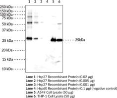
Heat shock protein 27 (Hsp27), also known as heat shock protein beta-1 (HspB1), is a member of the small heat shock protein (sHSP) family that is upregulated during conditions of cellular stress including heat shock, radiation, hypoxia, and exposure to reactive oxygen species (ROS).{40293,40294} It is composed of an N-terminal domain, a highly conserved alpha-crystallin domain, and a C-terminal domain. Hsp27 functions as a molecular chaperone to prevent protein aggregation in an ATPase-independent manner. This chaperone activity is altered by changes in oligomerization state or by post-translational modifications including phosphorylation at serine residues 15 and 82, which increases affinity for damaged polypeptides in response to heat shock.{40754} Hsp27 also works in complex with other chaperone proteins, such as Hsp70 (Item Nos. 22739 | 23002), to correct misfolded proteins. This protein also plays a role in apoptosis, proteasome activation, cell differentiation, and has been shown to interact with actin and intermediate filaments.{40295,40296,40297} Mutations in HSPB1 have been linked to hereditary neuromuscular diseases and cause Charcot-Marie-Tooth Disease Type 2 (CMT-2).{40298} Cayman’s Hsp27 (HspB1) Monoclonal Antibody can be used for Western blot and ELISA applications. The antibody recognizes Hsp27 (HspB1) at 25 kDa from human samples.
Brand:CaymanSKU:25692 - 100 µgAvailable on backorder
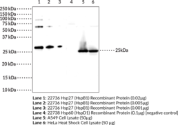
Heat shock protein 27 (Hsp27), also known as heat shock protein beta-1 (HspB1), is a member of the small heat shock protein (sHSP) family that is upregulated during conditions of cellular stress including heat shock, radiation, hypoxia, and exposure to reactive oxygen species (ROS).{40293,40294} It is composed of an N-terminal domain, a highly conserved alpha-crystallin domain, and a C-terminal domain. Hsp27 functions as a molecular chaperone to prevent protein aggregation in an ATPase-independent manner. This chaperone activity is altered by changes in oligomerization state or by post-translational modifications including phosphorylation at serine residues 15 and 82, which increases affinity for damaged polypeptides in response to heat shock.{40754} Hsp27 also works in complex with other chaperone proteins, such as Hsp70 (Item Nos. 22739 | 23002), to correct misfolded proteins. This protein also plays a role in apoptosis, proteasome activation, cell differentiation, and has been shown to interact with actin and intermediate filaments.{40295,40296,40297} Mutations in HSPB1 have been linked to hereditary neuromuscular diseases and cause Charcot-Marie-Tooth Disease Type 2 (CMT-2).{40298} Cayman’s Hsp27 (HspB1) Polyclonal Antibody can be used for ELISA, Immunofluorescence, Immunohistochemistry, and Western blot applications. The antibody recognizes Hsp27 (HspB1) at 25 kDa from human samples.
Brand:CaymanSKU:24530 - 300 µgAvailable on backorder
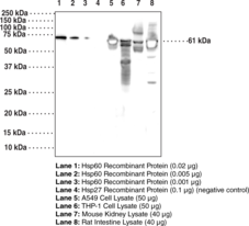
Heat shock protein 60 (Hsp60), also known as heat shock protein family D member 1 (HspD1), is an approximately 60 kDa protein that functions as a molecular chaperone.{35046} It belongs to the type I subclass of chaperonins and is found in eubacteria, mitochondria, and chloroplasts where its expression is induced by stress. Hsp60 primarily exists as a heptameric ring that it is converted to a tetradecameric double-ring structure in the presence of ATP.{36472} Within mitochondria, it associates with its co-chaperone, Hsp10, to form a barrel-like structure and refold proteins that have been shuttled to the mitochondria in an ATP-dependent manner.{35047,36472} Hsp60 also has extramitochondrial functions such as the production of proinflammatory cytokines in human leukocytes and activation of innate immune receptors.{35048,36473} Hsp60 expression is increased in the serum and saliva of patients with type 2 diabetes mellitus and mutations in HSPD1 lead to neurodegenerative diseases.{35049,36473} Cayman’s Hsp60 (HspD1) Monoclonal Antibody can be used for ELISA, IHC, and WB applications. The antibody recognizes Hsp60 (HspD1) at 61 kDa from human, mouse, and rat samples.
Brand:CaymanSKU:25693 - 100 µgAvailable on backorder

Heat shock protein 60 (Hsp60), also known as heat shock protein family D member 1 (HspD1), is an approximately 60 kDa protein that functions as a molecular chaperone.{35046} It belongs to the type I subclass of chaperonins and is found in eubacteria, mitochondria, and chloroplasts where its expression is induced by stress. Hsp60 primarily exists as a heptameric ring that it is converted to a tetradecameric double-ring structure in the presence of ATP.{36472} Within mitochondria, it associates with its co-chaperone, Hsp10, to form a barrel-like structure and refold proteins that have been shuttled to the mitochondria in an ATP-dependent manner.{36472,35047} Hsp60 also has extramitochondrial functions such as the production of proinflammatory cytokines in human leukocytes and activation of innate immune receptors.{35048,36473} Hsp60 expression is increased in the serum and saliva of patients with type 2 diabetes mellitus and mutations in HSPD1 lead to neurodegenerative diseases.{36473,35049} Cayman’s Hsp60 (HspD1) Polyclonal Antibody can be used for Western blot and ELISA applications. The antibody recognizes Hsp60 (HspD1) at 60 kDa from human samples.
Brand:CaymanSKU:24531 - 100 µgAvailable on backorder
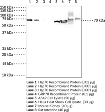
Heat shock protein 70s (Hsp70s) are abundant and stress-inducible 70 kDa molecular chaperone proteins encoded by a highly conserved, multigene family.{15480} They are monomeric proteins that can be divided into two functional domains: an N-terminal ATPase domain and a substrate binding domain that contains a highly conserved EEVD motif at its C-terminus. Hsp70s are found in the cytosol, nuclei, endoplasmic reticulum, mitochondria, and chloroplasts of eukaryotes, as well as in bacteria. They function as molecular chaperones that assist in a wide range of cellular processes, including refolding of aggregated or misfolded proteins, co- and post-translational folding and assembly of nascent peptides, membrane translocation of secretory and organellar proteins, controlling activity of regulatory nuclear receptors, kinases and transcription factors, as well as cooperativity with the Hsp90 chaperone system in eukaryotes.{36132} The Hsp70 chaperone cycle is ATP-dependent and initiated by transient interaction of the Hsp70 substrate binding domain with hydrophobic regions within a peptide or protein. It consists of an alteration between the low-affinity ATP-bound state with fast rates of substrate exchange and the high-affinity ADP bound state with slow rates of substrate exchange. Hsp70s are subject to a variety of post-translational modifications and their expression is upregulated under conditions of cellular stress and in a variety of disease states. Cayman’s Hsp70 (HspA1A) Monoclonal Antibody (Clone 7F6) can be used for Western blot, ELISA, immunohistochemistry, and immunoprecipitation applications. The antibody recognizes Hsp70, also known as HspA1A, at ~70 kDa from human and mouse samples.
Brand:CaymanSKU:25694 - 100 µgAvailable on backorder
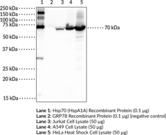
Heat shock protein 70s (Hsp70s) are abundant and stress-inducible 70 kDa molecular chaperone proteins encoded by a highly conserved, multigene family.{15480} They are monomeric proteins that can be divided into two functional domains: an N-terminal ATPase domain and a substrate binding domain that contains a highly conserved EEVD motif at its C-terminus. Hsp70s are found in the cytosol, nuclei, endoplasmic reticulum, mitochondria, and chloroplasts of eukaryotes, as well as in bacteria. They function as molecular chaperones that assist in a wide range of cellular processes, including refolding of aggregated or misfolded proteins, co- and post-translational folding and assembly of nascent peptides, membrane translocation of secretory and organellar proteins, controlling activity of regulatory nuclear receptors, kinases and transcription factors, as well as cooperativity with the Hsp90 chaperone system in eukaryotes.{36132} The Hsp70 chaperone cycle is ATP-dependent and initiated by transient interaction of the Hsp70 substrate binding domain with hydrophobic regions within a peptide or protein. It consists of an alteration between the low-affinity ATP-bound state with fast rates of substrate exchange and the high-affinity ADP bound state with slow rates of substrate exchange. Hsp70s are subject to a variety of post-translational modifications and their expression is upregulated under conditions of cellular stress and in a variety of disease states. Cayman’s Hsp70 (HspA1A) Polyclonal Antibody can be used for ELISA, Immunofluorescence, Immunohistochemistry, and Western blot applications. The antibody recognizes Hsp70, also known as HspA1A, at ~70 kDa from human samples.
Brand:CaymanSKU:24532 - 200 µgAvailable on backorder
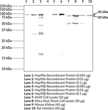
Hsp90 is a multidomain protein that functions as a molecular chaperone to assist in folding and activation of nascent peptides, refolding unfolded or misfolded proteins, and preventing protein aggregation.{15502} Hsp90α is the inducible cytosolic isoform of Hsp90 while Hsp90β is the constitutively active cytosolic isoform. Hsp90α and β are encoded by HSP90AA and HSP90AB, respectively in humans.{17930} C-terminal dimerization of Hsp90, coupled with ATPase molecular clamp activity induces a conformational change in the N-terminal nucleotide binding domain that facilitates substrate binding and initiates the chaperone cycle.{17932} Hsp90 interacts with many co-chaperones during its chaperone cycle including p23 and Sba1, which help recruit substrates to the Hsp90 complex, Hsp70 (Item Nos. 22739 | 23002), which loads nascent polypeptides onto the Hsp90 dimer, and the ATPase activator Aha1 that promotes ATP hydrolysis and substrate release.{17931,41851} Hsp90 is overexpressed in cancer cells and stabilizes client proteins that promote oncogenesis, including transcription factors, signaling proteins, and kinases.{17930,41851} Hsp90 also decreases α-synuclein fibril formation and toxicity as well as Q35 aggregation in in vitro models of Parkinson’s and Huntington’s disease, respectively, implying a role in neurodegenerative disease.{41852} Cayman’s Hsp90α/β Polyclonal Antibody can be used for Western blot and ELISA applications. This antibody recognizes Hsp90α at 85 kDa and Hsp90β at 83 kDa from human, mouse, and rat samples.
Brand:CaymanSKU:24559 - 500 µlAvailable on backorder
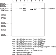
Heat shock protein 90 α (Hsp90α) is the inducible cytosolic isoform of Hsp90 that is encoded by HSP90AA in humans.{17930} Hsp90 is a multidomain protein that functions as a molecular chaperone to assist in folding and activation of nascent peptides, refolding unfolded or misfolded proteins, and preventing protein aggregation.{15502} C-terminal dimerization of Hsp90, coupled with ATPase molecular clamp activity induces a conformational change in the N-terminal nucleotide binding domain that facilitates substrate binding and initiates the chaperone cycle.{17932} Hsp90 interacts with many co-chaperones during its chaperone cycle including p23 and Sba1, which help recruit substrates to the Hsp90 complex, Hsp70 (Item Nos. 22739 | 23002), which loads nascent polypeptides onto the Hsp90 dimer, and the ATPase activator Aha1 that promotes ATP hydrolysis and substrate release.{17931,41851} Hsp90 is overexpressed in cancer cells and stabilizes client proteins that promote oncogenesis, including transcription factors, signaling proteins, and kinases.{17930,41851} Hsp90 also decreases α-synuclein fibril formation and toxicity as well as Q35 aggregation in in vitro models of Parkinson’s and Huntington’s disease, respectively, implying a role in neurodegenerative disease.{41852} Cayman’s Hsp90α Polyclonal Antibody can be used for Western blot and ELISA applications. This antibody recognizes Hsp90α at 85 kDa from human, mouse, and rat samples.
Brand:CaymanSKU:25725 - 500 µlAvailable on backorder
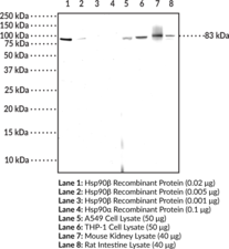
Heat shock protein 90 β (Hsp90β) is the constitutively active cytosolic isoform of Hsp90 that is encoded by HSP90AB in humans.{17930} Hsp90 is a multidomain protein that functions as a molecular chaperone to assist in folding and activation of nascent peptides, refolding unfolded or misfolded proteins, and preventing protein aggregation.{15502} C-terminal dimerization of Hsp90, coupled with ATPase molecular clamp activity induces a conformational change in the N-terminal nucleotide binding domain that facilitates substrate binding and initiates the chaperone cycle.{17932} Hsp90 interacts with many co-chaperones during its chaperone cycle including p23 and Sba1, which help recruit substrates to the Hsp90 complex, Hsp70 (Item Nos. 22739 | 23002), which loads nascent polypeptides onto the Hsp90 dimer, and the ATPase activator Aha1 that promotes ATP hydrolysis and substrate release.{17931,41851} Hsp90 is overexpressed in cancer cells and stabilizes client proteins that promote oncogenesis, including transcription factors, signaling proteins, and kinases.{17930,41851} Hsp90 also decreases α-synuclein fibril formation and toxicity as well as Q35 aggregation in in vitro models of Parkinson’s and Huntington’s disease, respectively, implying a role in neurodegenerative disease.{41852} Cayman’s Hsp90β Monoclonal Antibody (Clone8D6) can be used for Western blot and ELISA applications. This antibody recognizes Hsp90β at 83 kDa from human, mouse, and rat samples.
Brand:CaymanSKU:25695 - 100 µgAvailable on backorder
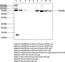
Heat shock protein 90 β (Hsp90β) is the constitutively active cytosolic isoform of Hsp90 that is encoded by HSP90AB in humans.{17930} Hsp90 is a multidomain protein that functions as a molecular chaperone to assist in folding and activation of nascent peptides, refolding unfolded or misfolded proteins, and preventing protein aggregation.{15502} C-terminal dimerization of Hsp90, coupled with ATPase molecular clamp activity induces a conformational change in the N-terminal nucleotide binding domain that facilitates substrate binding and initiates the chaperone cycle.{17932} Hsp90 interacts with many co-chaperones during its chaperone cycle including p23 and Sba1, which help recruit substrates to the Hsp90 complex, Hsp70 (Item Nos. 22739 | 23002), which loads nascent polypeptides onto the Hsp90 dimer, and the ATPase activator Aha1 that promotes ATP hydrolysis and substrate release.{17931,41851} Hsp90 is overexpressed in cancer cells and stabilizes client proteins that promote oncogenesis, including transcription factors, signaling proteins, and kinases.{17930,41851} Hsp90 also decreases α-synuclein fibril formation and toxicity as well as Q35 aggregation in in vitro models of Parkinson’s and Huntington’s disease, respectively, implying a role in neurodegenerative disease.{41852} Cayman’s Hsp90β Polyclonal Antibody can be used for Western blot, ELISA, IHC, and IF applications. This antibody recognizes Hsp90β at 83 kDa from human, mouse, and rat samples.
Brand:CaymanSKU:25730 - 500 µLAvailable on backorder
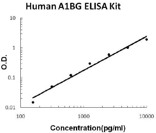
For quantitative detection of human A1BG in cell culture supernates, cell lysates, serum and plasma (heparin, EDTA).
Brand:Boster BioSKU:EK1488Available on backorder
For quantitative detection of human A2M in cell culture supernates, cell lysates, serum and plasma (heparin, EDTA).
Brand:Boster BioSKU:EK1118Available on backorder
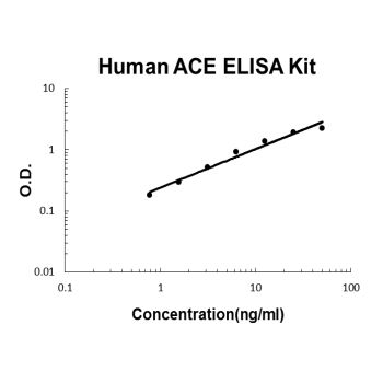
For quantitative detection of human ACE in cell culture supernates, serum, plasma(heparin) and saliva.
Brand:Boster BioSKU:EK0557Available on backorder
Brand:Boster BioSKU:EK0997Available on backorder
For quantitative detection of human Activin A in cell culture supernates, serum, plasma(heparin, EDTA) and saliva.
Brand:Boster BioSKU:EK0301Available on backorder
Brand:Boster BioSKU:EK1446Available on backorder
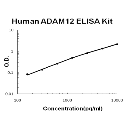
For quantitative detection of human ADAM12 in cell culture supernates, serum, plasma(heparin) and urine.
Brand:Boster BioSKU:EK0934Available on backorder
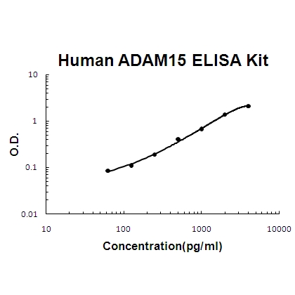
For quantitative detection of human ADAM15 in cell culture supernates, cell lysates, serum and plasma (heparin, EDTA).
Brand:Boster BioSKU:EK1484Available on backorder
 Brand:Boster BioSKU:EK0650
Available on backorder
 Brand:Boster BioSKU:EK1341
Available on backorder
Brand:Boster BioSKU:EK1340Available on backorder
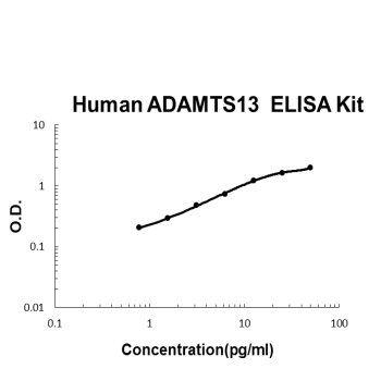
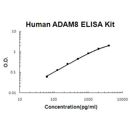
For quantitative detection of human ADAMTS13 in cell culture supernates, serum and plasma(heparin, EDTA, citrate).
Brand:Boster BioSKU:EK0927Available on backorder
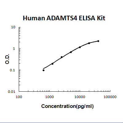
For quantitative detection of human ADAMTS4 in cell culture supernates, cell lysates, serum and plasma (heparin, EDTA).
Brand:Boster BioSKU:EK1372Available on backorder
For quantitative detection of human Adiponectin in cell culture supernates, serum, plasma(heparin, EDTA)and urine.
Brand:Boster BioSKU:EK0595Available on backorder
The Fast version of Picokine ELISA kits, assay takes less than 1.5 hours. Detect Human Adiponectin/ADIPOQ with <60pg/ml sensitivity. Format: 96-well plate with removable strips. Compatible samples: cell culture supernates, serum, plasma(heparin, EDTA)and urine. This is a TMB colorimetric sandwich ELISA kit with short assay time and fast experiment set up. Adiponectin/ADIPOQ tissue specificity:
Brand:Boster BioSKU:FEK0595Available on backorder
 Brand:Boster BioSKU:EK1487
Available on backorder
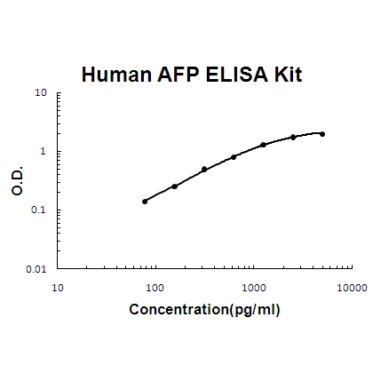
For quantitative detection of human AFP in cell culture supernates, cell lysates, serum and plasma (heparin, EDTA).
Brand:Boster BioSKU:EK0651Available on backorder
 Brand:Boster BioSKU:EK0909
Available on backorder
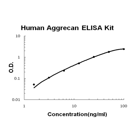
The Fast version of Picokine ELISA kits, assay takes less than 1.5 hours. Detect Human Aggrecan/ACAN with <50pg/ml sensitivity. Format: 96-well plate with removable strips. Compatible samples: cell culture supernates, serum and urine. This is a TMB colorimetric sandwich ELISA kit with short assay time and fast experiment set up. Aggrecan/ACAN tissue specificity: Restricted to cartilages. .
Brand:Boster BioSKU:FEK0909Available on backorder
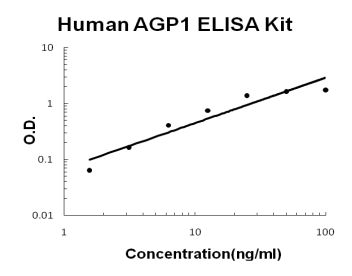
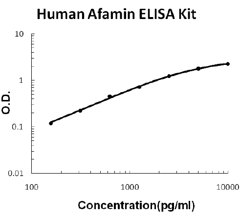
For quantitative detection of human AGP1 in cell culture supernates, cell lysates, serum and plasma (heparin, EDTA).
Brand:Boster BioSKU:EK1486Available on backorder
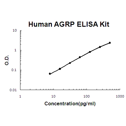
For quantitative detection of human AGRP in cell culture supernates, serum, plasma (heparin, EDTA, citrate) and urine.
Brand:Boster BioSKU:EK1427Available on backorder
 Brand:Boster BioSKU:EK1465
Available on backorder
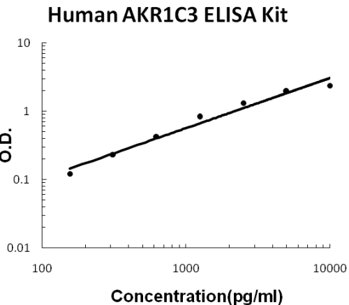
For quantitative detection of human AKR1C3 in cell culture supernates, serum and plasma (heparin, EDTA).
Brand:Boster BioSKU:EK1869Available on backorder
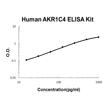
For quantitative detection of human AKR1C4 in cell culture supernates, serum and plasma (heparin, EDTA).
Brand:Boster BioSKU:EK1870Available on backorder
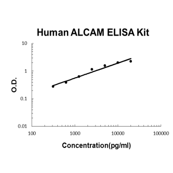
For quantitative detection of human ALCAM in cell culture supernates, cell lysates, serum and plasma (heparin, EDTA).
Brand:Boster BioSKU:EK0995Available on backorder
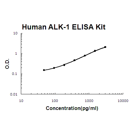
For quantitative detection of human ALK-1 in cell culture supernates, cell lysates, serum and plasma (heparin, EDTA).
Brand:Boster BioSKU:EK1518Available on backorder
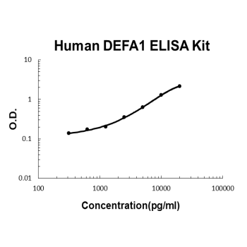
For quantitative detection of human DEFA1 in cell culture supernates, serum and plasma (heparin, EDTA).
Brand:Boster BioSKU:EK1514Available on backorder
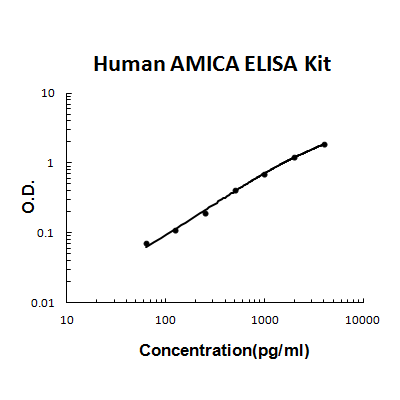
For quantitative detection of human AMICA/JAML in cell culture supernates, serum and plasma (heparin, EDTA).
Brand:Boster BioSKU:EK1730Available on backorder

For quantitative detection of human Amphiregulin in cell culture supernates, serum, plasma(heparin, EDTA), saliva and human milk.
Brand:Boster BioSKU:EK0304Available on backorder
For quantitative detection of human ANG in cell culture supernates, cell lysates, serum and plasma (heparin, EDTA).
Brand:Boster BioSKU:EK0305Available on backorder
The Fast version of Picokine ELISA kits, assay takes less than 1.5 hours. Detect Human Angiogenin/ANG with <12pg/ml sensitivity. Format: 96-well plate with removable strips. Compatible samples: cell culture supernates, cell lysates, serum and plasma (heparin, EDTA). This is a TMB colorimetric sandwich ELISA kit with short assay time and fast experiment set up. Angiogenin/ANG tissue specificity: Expressed predominantly in the liver. Also The Fast version of Picokine ELISA kits, assay takes less than 1.5 hours. Detected in endothelial cells and spinal cord neurons. .
Brand:Boster BioSKU:FEK0559Available on backorder
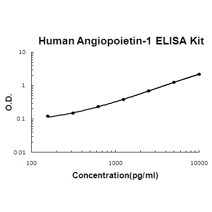
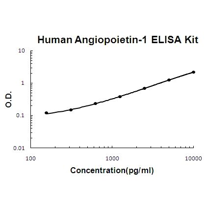
For quantitative detection of human Angiopoietin-1 in cell culture supernates, serum, plasma(heparin, EDTA), saliva and human milk.
Brand:Boster BioSKU:EK0559Available on backorder
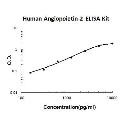
For quantitative detection of human Angiopoietin-2 in cell culture supernates, serum, plasma(heparin, EDTA), saliva and human milk.
Brand:Boster BioSKU:EK0657Available on backorder
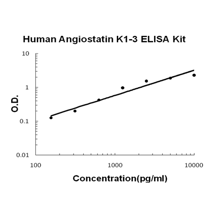
For quantitative detection of human Angiostatin K1-3 in cell culture supernates, serum and plasma(heparin, EDTA).
Brand:Boster BioSKU:EK0905Available on backorder
The Fast version of Picokine ELISA kits, assay takes less than 1.5 hours. Detect Human Plasminogen/PLG with <10pg/ml sensitivity. Format: 96-well plate with removable strips. Compatible samples: serum, plasma and cell culture supernates. This is a TMB colorimetric sandwich ELISA kit with short assay time and fast experiment set up. Plasminogen/PLG tissue specificity: Present in plasma and many other extracellular fluids. It is synthesized in the liver.
Brand:Boster BioSKU:FEK0905Available on backorder
For quantitative detection of human ANGPTL2 in cell culture supernates, serum and plasma (heparin, EDTA).
Brand:Boster BioSKU:EK2039Available on backorder
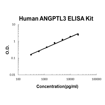
For quantitative detection of human ANGPTL3 in cell culture supernates, cell lysates, serum and plasma (heparin, EDTA).
Brand:Boster BioSKU:EK1374Available on backorder
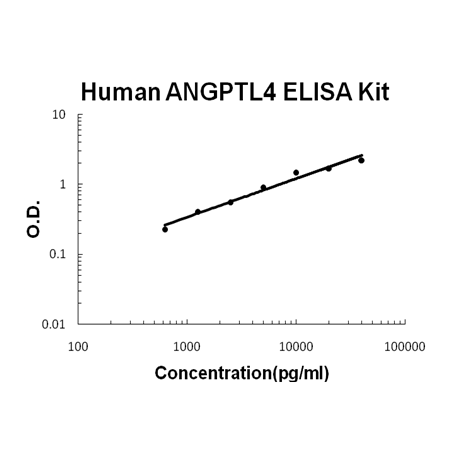
For quantitative detection of human ANGPTL4 in cell culture supernates, cell lysates, serum and plasma (heparin, EDTA).
Brand:Boster BioSKU:EK0960Available on backorder
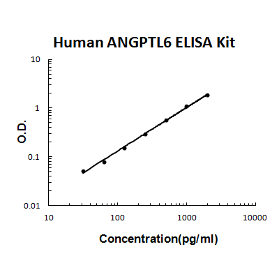
For quantitative detection of human ANGPTL6 in cell culture supernates, serum and plasma (heparin, EDTA).
Brand:Boster BioSKU:EK1993Available on backorder
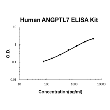
For quantitative detection of human ANGPTL7 in cell culture supernates, serum and plasma (heparin, EDTA).
Brand:Boster BioSKU:EK2035Available on backorder
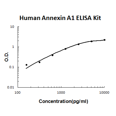
For quantitative detection of human Annexin A1 in cell culture supernates, serum and plasma (heparin, EDTA).
Brand:Boster BioSKU:EK1745Available on backorder
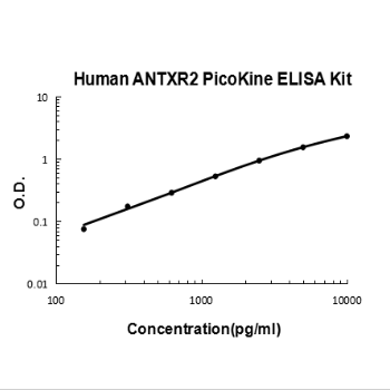
For quantitative detection of human ANTXR2 in cell culture supernates, serum and plasma (heparin, EDTA, citrate).
Brand:Boster BioSKU:EK2108Available on backorder
For quantitative detection of human APLP1 in cell culture supernates, serum and plasma (heparin, EDTA).
Brand:Boster BioSKU:EK2103Available on backorder
For quantitative detection of human APOA1 in cell culture supernates, serum, plasma(heparin, EDTA) and urine.
Brand:Boster BioSKU:EK1456Available on backorder
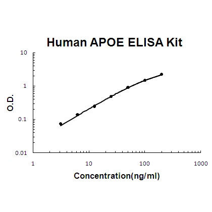
For quantitative detection of human APOE in cell culture supernates, serum, plasma(heparin, EDTA), saliva, milk and urine.
Brand:Boster BioSKU:EK1455Available on backorder
For quantitative detection of human APOH in cell culture supernates, serum and plasma (heparin, EDTA).
Brand:Boster BioSKU:EK2026Available on backorder
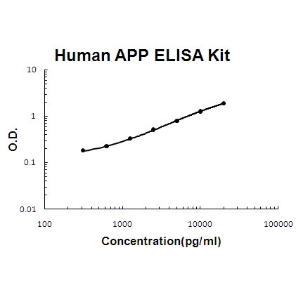
For quantitative detection of human APP in cell culture supernates, cell lysates, serum and plasma (heparin, EDTA).
Brand:Boster BioSKU:EK0658Available on backorder
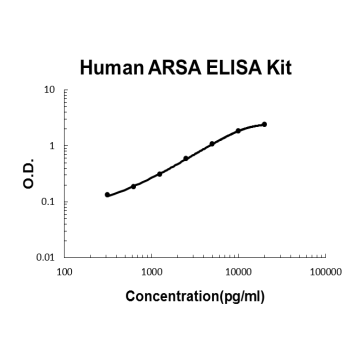
For quantitative detection of human ARSA in cell culture supernates, serum and plasma (heparin, EDTA, citrate).
Brand:Boster BioSKU:EK2077Available on backorder
For quantitative detection of human ARSB in cell culture supernates, serum and plasma (heparin, EDTA, citrate).
Brand:Boster BioSKU:EK2078Available on backorder
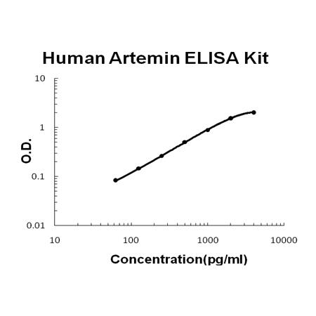
For Quantitative Detection of human Artemin in cell culture supernates, serum and plasma (heparin, EDTA).
Brand:Boster BioSKU:EK1534Available on backorder
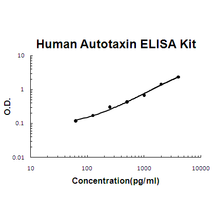
For Quantitative Detection of human Autotaxin in cell culture supernates, serum, plasma (heparin), urine and human milk.
Brand:Boster BioSKU:EK1655Available on backorder
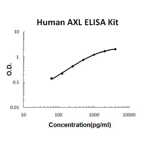
For quantitative detection of human AXL in cell culture supernates, cell lysates, serum and plasma (heparin, EDTA).
Brand:Boster BioSKU:EK0659Available on backorder
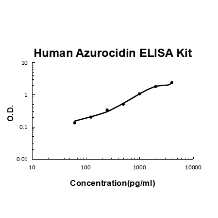
For quantitative detection of human Azurocidin in cell culture supernates, cell lysates, serum and plasma (heparin, EDTA).
Brand:Boster BioSKU:EK1161Available on backorder
For quantitative detection of human B2M in cell culture supernates, serum, plasma (heparin, EDTA), saliva, urine and human milk.
Brand:Boster BioSKU:EK1691Available on backorder
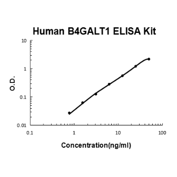
For quantitative detection of human B4GALT1 in cell culture supernates, serum and plasma (heparin, EDTA).
Brand:Boster BioSKU:EK1880Available on backorder
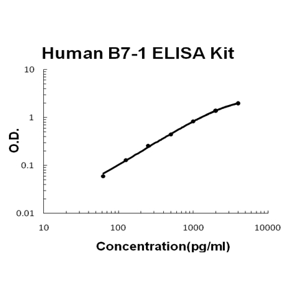
 Brand:Boster BioSKU:EK0707
Available on backorder
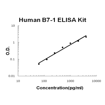
The Fast version of Picokine ELISA kits, assay takes less than 1.5 hours. Detect Human B7-1/CD80 with <10pg/ml sensitivity. Format: 96-well plate with removable strips. Compatible samples: cell culture supernates and serum. This is a TMB colorimetric sandwich ELISA kit with short assay time and fast experiment set up. B7-1/CD80 tissue specificity: Expressed on activated B-cells, macrophages and dendritic cells.
Brand:Boster BioSKU:FEK0707Available on backorder
For quantitative detection of human BAFF in cell culture supernates, cell lysates, serum and plasma (heparin, EDTA).
Brand:Boster BioSKU:EK0663Available on backorder
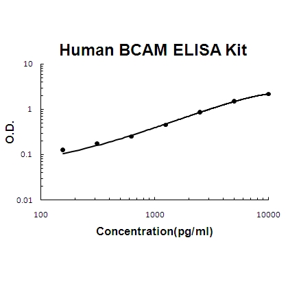
For quantitative detection of human BCAM in cell culture supernates, cell lysates, serum and plasma (heparin, EDTA).
Brand:Boster BioSKU:EK1546Available on backorder
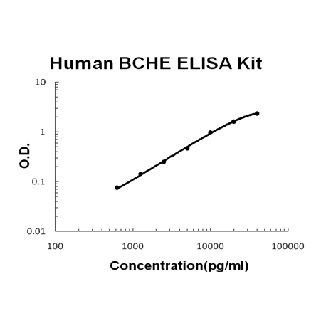
For quantitative detection of human BCHE in cell culture supernates, serum, plasma (heparin, EDTA), saliva, urine and human milk.
Brand:Boster BioSKU:EK1507Available on backorder
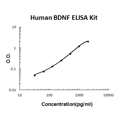
For quantitative detection of human BDNF in cell culture supernates, cell lysates, serum and plasma (heparin, EDTA, citrate).
Brand:Boster BioSKU:EK0307Available on backorder
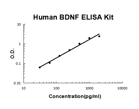
For the quantitation of Human BDNF concentrations in cell culture supernates, cell lysates, serum and plasma (heparin, EDTA, citrate).
Brand:Boster BioSKU:FEK0307Available on backorder
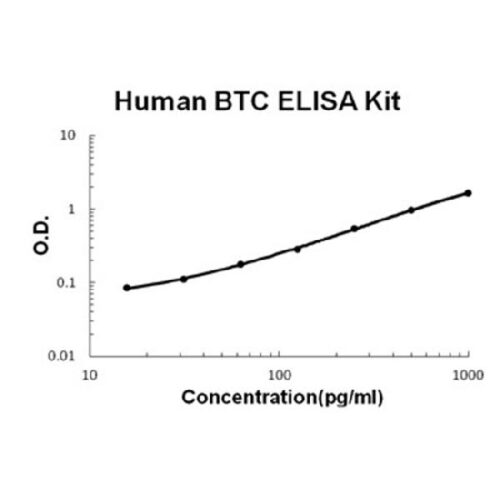
For quantitative detection of human Betacellulin in cell culture supernates, cell lysates, serum and plasma (heparin, EDTA).
Brand:Boster BioSKU:EK0980Available on backorder
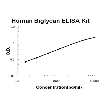
For quantitative detection of human Biglycan in cell culture supernates, serum and plasma(heparin, EDTA).
Brand:Boster BioSKU:EK1357Available on backorder
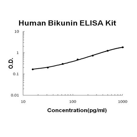
For Quantitative Detection of human Bikunin in cell culture supernates, serum and plasma (heparin, EDTA).
Brand:Boster BioSKU:EK1598Available on backorder