ELISA Kits
Showing 751–900 of 3623 results
-
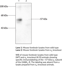
GABAA receptors are ligand-gated chloride channels that mediate the effects of the inhibitory neurotransmitter GABA in the CNS.{46949,46950} They are postsynaptic heteropentameric receptors that contains protein subunits from the following isoforms: α1-6, β1-4, γ1-3, δ, ε, π, θ, and ρ1-3, arranged around a central pore. Phasic inhibitory synaptic transmission is regulated by α1β2γ2 subunit-containing GABAA receptors, the major isoform found in the brain.{46950,46951} The α subunit of GABAA receptors interfaces with a β subunit to form the GABA binding site that initiates GABA-induced action potentials and forms the benzodiazepine binding site with the γ subunit. The GABAA receptor α6 subunit is expressed in granule cells of the cerebellum and cochlear nucleus.{46947} Expression of GABRA6, which encodes the α6 subunit isoform, is decreased in withdrawal seizure-prone, but not withdrawal seizure-resistant, mice after chronic ethanol administration. Point mutation of the arginine residue at position 100 (R100Q) of the GABAA receptor α6 subunit increases sensitivity to diazepam and ethanol in rats and humans.{46948} Cayman’s GABAA Receptor α6 Subunit Polyclonal Antibody can be used for Western blot (WB) applications. The antibody recognizes the GABAA receptor α6 subunit at approximately 57 kDa from mouse and rat samples.
Brand:CaymanSKU:29272 - 100 µlAvailable on backorder
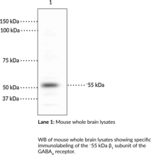
GABAA receptors are ligand-gated chloride channels that mediate the effects of the inhibitory neurotransmitter GABA in the CNS.{46949,46950} They are postsynaptic heteropentameric receptors that contain protein subunits from the following isoforms: α1-6, β1-4, γ1-3, δ, ε, π, θ, and ρ1-3, arranged around a central pore. Phasic inhibitory synaptic transmission is regulated by α1β2γ2 subunit-containing GABAA receptors, the major isoform found in the brain.{46950,46951} The β subunit of GABAA receptors interfaces with an α subunit to form the GABA binding site that initiates GABA-induced action potentials and forms the benzodiazepine binding site with the γ subunit. Phosphorylation of the β1 subunit by PKA or PKC inhibits binding of the β1 subunit with the clathrin adaptor protein AP2 and reduces GABAA receptor endocytosis.{55097} Mice expressing mutations in Gabrb1, which encodes the β1 subunit isoform, have increased GABAA receptor-mediated tonic inhibition in the nucleus accumbens and increased alcohol consumption compared to wild-type mice.{55212} SNPs in GABRB1 are associated with increased impulsivity and reward sensitivity in human adolescents.{55217} Cayman’s GABAA Receptor β1 Polyclonal Antibody can be used for Western blot (WB) applications. The antibody recognizes the GABAA receptor β1 subunit at approximately 55 kDa from mouse and rat samples.
Brand:CaymanSKU:29273 - 100 µlAvailable on backorder
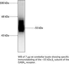
GABAA receptors are ligand-gated chloride channels that mediate the effects of the inhibitory neurotransmitter GABA in the CNS.{46949,46950} They are postsynaptic heteropentameric receptors that contain protein subunits from the following isoforms: α1-6, β1-4, γ1-3, δ, ε, π, θ, and ρ1-3, arranged around a central pore. Phasic inhibitory synaptic transmission is regulated by α1β2γ2 subunit-containing GABAA receptors, the major isoform found in the brain.{46950,46951} The β subunit of GABAA receptors interfaces with an α subunit to form the GABA binding site that initiates GABA-induced action potentials and forms the benzodiazepine binding site with the γ subunit. Phosphorylation of the β2 subunit by PKC inhibits binding of the β2 subunit with the clathrin adaptor protein AP2 and reduces GABAA receptor endocytosis.{55097} SNPs in GABRB2, which encodes the β2 subunit isoform, have been found in patients with schizophrenia.{55096} Cayman’s GABAA Receptor β2 Polyclonal Antibody can be used for Western blot (WB) applications. The antibody recognizes the GABAA receptor β2 subunit at approximately 55 kDa from mouse and rat samples.
Brand:CaymanSKU:29274 - 100 µlAvailable on backorder
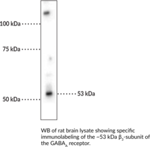
GABAA receptors are ligand-gated chloride channels that mediate the effects of the inhibitory neurotransmitter GABA in the CNS.{46949,46950} They are postsynaptic heteropentameric receptors that contain protein subunits from the following isoforms: α1-6, β1-4, γ1-3, δ, ε, π, θ, and ρ1-3, arranged around a central pore. Phasic inhibitory synaptic transmission is regulated by α1β2γ2 subunit-containing GABAA receptors, the major isoform found in the brain.{46950,46951} The β subunit of GABAA receptors interfaces with an α subunit to form the GABA binding site that initiates GABA-induced action potentials and forms the benzodiazepine binding site with the γ subunit. β3 subunit-containing GABAA receptors are widely expressed in the cerebral cortex, cerebellum, olfactory bulb, and hippocampus.{55098} Phosphorylation of the β3 subunit by PKA or PKC inhibits binding of the β3 subunit with the clathrin adaptor protein AP2 and reduces GABAA receptor endocytosis.{55097} Mutations in GABRB3, which encodes the β3 subunit isoform, have been found in patients with childhood absence epilepsy (CAE), infantile spasms (IS), and Lennox-Gastaut syndrome (LGS).{46950} Cayman’s GABAA Receptor β3 Subunit Polyclonal Antibody can be used for immunohistochemistry (IHC) and Western blot (WB) applications. The antibody recognizes the GABAA receptor β3 subunit at approximately 53 kDa from rat and mouse samples.
Brand:CaymanSKU:29275 - 100 µlAvailable on backorder
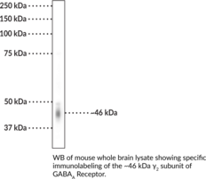
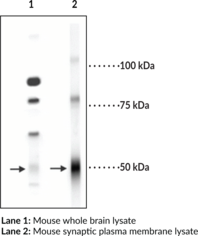
GABAA receptors are ligand-gated chloride channels that mediate the effects of the inhibitory neurotransmitter GABA in the CNS.{46949,46950} They are postsynaptic heteropentameric receptors that contain protein subunits from the following isoforms: α1-6, β1-4, γ1-3, δ, ε, π, θ, and ρ1-3, arranged around a central pore. Phasic inhibitory synaptic transmission is regulated by α1β2γ2 subunit-containing GABAA receptors, the major isoform found in the brain.{46950,46951} The γ2 subunit is the most abundant subunit in the brain and is required for clustering and postsynaptic localization of α1 or α2 subunit-containing GABAA receptors in mouse brain.{56129} Mutations in GABRG2, the gene encoding the γ2 subunit, have been found in patients with childhood absence epilepsy (CAE), genetic epilepsy with febrile seizures plus (GEFS+), Dravet syndrome, and idiopathic genetic generalized epilepsy (GGE).{46950} Cayman’s GABAA Receptor γ2 Subunit Polyclonal Antibody can be used for immunohistochemistry (IHC) and Western blot (WB) applications. The antibody recognizes the GABAA receptor γ2 subunit at approximately 46 kDa from mouse and rat samples.
Brand:CaymanSKU:29276 - 100 µlAvailable on backorder
Gamma-aminobutyric acid (GABA) is the primary inhibitory neurotransmitter in the central nervous system, causing a hyperpolarization of the membrane through the opening of a Cl- channel associated with the GABAA receptor (GABAA-R) subtype. GABAA-Rs are important therapeutic targets for a range of sedative, anxiolytic, and hypnotic agents and are implicated in several diseases including epilepsy, anxiety, depression, and substance abuse. The GABAA-R is a multimeric subunit complex. To date six α’s, four β’s, and four γ’s, plus alternative splicing variants of some of these subunits, have been identified. Injection in oocytes or mammalian cell lines of cRNA coding for α and β subunits results in the expression of functional GABAA-Rs sensitive to GABA. However, co-expression of a γ subunit is required for benzodiazepine modulation. The various effects of the benzodiazepines in brain may also be mediated via different α subunits of the receptor. More recently there have been a number of studies demonstrating that the δ subunit of the receptor may affect subunit assembly and may also confer differential sensitivity to neurosteroids and to ethanol.
Brand:CaymanSKU:10600 - 1 eaAvailable on backorder

This protein is a positive regulator for the gene expression of the galactose-induced genes such as GAL1, GAL2, GAL7, GAL10, and MEL1 which code for the enzymes used to convert galactose to glucose. [Bertin Catalog No. G01015]
Brand:CaymanSKU:32784 - 100 µlAvailable on backorder

This protein is a positive regulator for the gene expression of the galactose-induced genes such as GAL1, GAL2, GAL7, GAL10, and MEL1 which code for the enzymes used to convert galactose to glucose. [Bertin Catalog No. G01017]
Brand:CaymanSKU:32786 - 100 µlAvailable on backorder
This protein is a positive regulator for the gene expression of the galactose-induced genes such as GAL1, GAL2, GAL7, GAL10, and MEL1 which code for the enzymes used to convert galactose to glucose. [Bertin Catalog No. G01018]
Brand:CaymanSKU:32787 - 100 µlAvailable on backorder
This protein is a positive regulator for the gene expression of the galactose-induced genes such as GAL1, GAL2, GAL7, GAL10, and MEL1 which code for the enzymes used to convert galactose to glucose. [Bertin Catalog No. G01016]
Brand:CaymanSKU:32785 - 100 µLAvailable on backorder

Ganglioside GD1b (Item No. 19569) is an acidic glycosphingolipid that contains two sialic acid residues linked to an inner galactose unit. It is a component of plasma membranes where it packs densely with cholesterol to form lipid microdomains that modulate both intra- and intercellular signaling events.{31218} The concentration of ganglioside GD1b in human brain increases with age, constituting 7.85% of total sialic acid in the brain of 0- to 10-year-old subjects and 20.29% in 11- to 30-year-old subjects.{31219} Ganglioside GD1b levels are positively correlated with pilocytic astrocytoma tumor grade, and GD1b has been detected in various other gliomas, including primitive neuroectodermal tumors, glioblastomas, and anaplastic astrocytomas.{54530} Ganglioside GD1b Polyclonal Antibody can be used for ELISA and TLC immunoblotting applications. [Matreya, LLC. Catalog No. 1964]
Brand:CaymanSKU:28642 - 50 µlAvailable on backorder
Ganglioside GD3 (Item No. 17481) is synthesized by the addition of two sialic acid residues to lactosylceramide and can serve as a precursor to the formation of more complex gangliosides by the action of glycosyl- and sialyltransferases.{31218} It induces apoptosis in HuT-78 cutaneous T cell lymphoma cells in a concentration-dependent manner and disrupts the mitochondrial membrane potential when used at a concentration of 200 μM.{5033} Expression of ganglioside GD3 in GD3-negative SK-MEL-28-N1 malignant melanoma cells increases both cell proliferation and invasion in vitro.{28470} Ganglioside GD3-deficient adult mice exhibit progressive loss of the neural stem cell (NSC) pool and impaired neurogenesis.{37782} Ganglioside GD3 Monoclonal Antibody can be used for ELISA and TLC immunobloting. [Matreya, LLC. Catalog No. 1977]
Brand:CaymanSKU:28644 - 50 µLAvailable on backorder
Ganglioside GM1 asialo (Item No. 15586) is a component of cellular lipid rafts and can be formed by the cleavage of the sialic acid residue from ganglioside GM1 (Item No. 19579) by neuraminidase.{25489,37774} Ganglioside GM1 asialo is a glycolipid receptor for P. aeruginosa flagellin and stimulates defensive responses in host cells, including extracellular ATP release, calcium mobilization, and ERK1/2 phosphorylation when stimulated by flagellin and an anti-ganglioside GM1 asialo antibody.{25488} The percentage of ganglioside GM1 asialo-positive natural killer (NK) and CD8+ T cells in the lung is increased in a mouse model of respiratory syncytial virus (RSV) infection compared with healthy animals.{25489} Depletion of ganglioside GM1 asialo-positive NK and T cells reduces IFN-γ levels in the lung, reduces weight loss, and increases lung viral load in RSV-infected mice. Ganglioside GM1 Asialo Polyclonal Antibody can be used for ELISA and TLC immunoblotting. [Matreya, LLC. Catalog No. 1950]
Brand:CaymanSKU:28638 - 100 µlAvailable on backorder
Ganglioside GM1 is a monosialylated ganglioside and the prototypic ganglioside for those containing one sialic acid residue.{31218,52660} It is found in a large variety of cells, including immune cells and neurons, and is enriched in lipid rafts in the cell membrane.{53951} It associates with growth factor receptors, including TrkA, TrkB, and the GDNF receptor complex containing Ret and GFRα, and is required for TrkA expression on the cell surface. Ganglioside GM1 interacts with other proteins to increase calcium influx, affecting various calcium-dependent processes, including inducing neuronal outgrowth during differentiation. Ganglioside GM1 acts as a receptor for cholera toxin, which binds to its oligosaccharide group, facilitating toxin cell entry into epithelial cells of the jejunum.{53952,53953} Similarly, it is bound by the heat-labile enterotoxin from E. coli in the pathogenesis of traveler’s diarrhea.{53954} Ganglioside GM1 sensitizes inactivated T cells to TNF-α-induced apoptosis and induces apoptosis of activated T cells even in the absence of TNF-α.{52850} Ganglioside GM1 is found at higher levels on T cells isolated from patients with renal cell carcinoma (RCC) compared with T cells from patients without cancer. Levels of ganglioside GM1 are decreased in the substantia nigra pars compacta in postmortem brain from patients with Parkinson’s disease.{53951} Ganglioside GM1 gangliosidosis, characterized by a deficiency in GM1-β-galactosidase, the enzyme that degrades ganglioside GM1, leads to accumulation of the gangliosides GM1 and GA1 in neurons and can be fatal in infants.{31218} [Matreya, LLC. Catalog No. 1954]
Brand:CaymanSKU:28640 - 100 µlAvailable on backorder
Ganglioside GM2 asialo (asialo- GM2; Item No. 24629) is a glycosphingolipid containing three monosaccharide residues and a fatty acid of variable chain length but lacking the sialic acid residue present on ganglioside M2. Asialo-GM2 levels are low-to-undetectable in normal human brain, but it accumulates in the brain of patients with Tay-Sachs and Sandhoff disease, which are neurodegenerative disorders characterized by deficiency of lysosomal β-hexosaminidase A and B, respectively.{40891} It also binds to various bacteria, including Pseudomonas isolates derived from cystic fibrosis patients.{40892} Ganglioside GM2 Asialo Polyclonal Antibody can be used for ELISA and TLC immunoblotting. [Matreya, LLC. Catalog No. 1951]
Brand:CaymanSKU:28639 - 50 µlAvailable on backorder
Gap-43 is thought to have an important role in development and plasticity because it is expressed at high levels in neuronal growth cones during development and during axonal regeneration.{14428} There is also evidence from knockout animals that Gap-43 serves to amplify path finding signals from the growth cone.{14431} Gap-43 is thought to mediate at least some of these effects via interaction with actin. Importantly, phosphorylation at Ser41 by protein kinase C (Catalog No. 1609-PKC) modulates the interaction of Gap-43 with actin and may also affect neurotransmitter release during forms of plasticity like LTP.{14429,14430}
Brand:CaymanSKU:10009400 - 1 eaAvailable on backorder
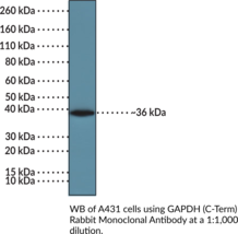
Glyceraldehyde-3-phosphate dehydrogenase (GAPDH) is an enzyme that catalyzes the conversion of glyceraldehyde-3-phosphate (Item No. 17865) to 1,3-bisphosphoglycerate during glycolysis and is involved in numerous additional cellular processes, including intracellular trafficking, receptor-mediated signaling, apoptosis, DNA repair, and the oxidative stress response.{54459,54460} It exists as a tetramer and is composed of an N-terminal domain, which contains binding sites for NAD+, phosphatidylserine, RNA, and glutathione, and a C-terminal catalytic domain.{54461} GAPDH is widely expressed and primarily localizes to the cytosol, where it has roles in glycolysis and intracellular trafficking.{54459,60077} It also localizes to the nucleus, mediating DNA integrity, gene transcription, and apoptosis, as well as to cellular membranes, where it has roles in membrane fusion and iron transport.{60077} GAPDH expression is increased by insulin, hypoxia-inducible factor-1 (HIF-1), p53, and nitric oxide (NO) and decreased by acetylated histones.{54459,54462} Aberrant mRNA and protein levels of GAPDH have been found in tumor biopsies from patients with a variety of cancers, including lung, renal cell, colorectal, or breast cancer.{60078} Cayman’s GAPDH (C-Term) Rabbit Monoclonal Antibody can be used for immunocytochemistry (ICC), immunoprecipitation (IP), chromatin immunoprecipitation (ChIP), and Western blot (WB) applications. The antibody recognizes the C-terminal region of GAPDH at approximately 36 kDa.
Brand:CaymanSKU:32128 - 100 µlAvailable on backorder
 Brand:CaymanSKU:32901 - 100 µl
Available on backorder
Glial fibrillary acidic protein (GFAP) is a 40-53 kDa monomeric molecule found only in adult glial cells of the central nervous system (CNS) and represents the major part of the cytoskeleton of astrocytes. Traumatic injury to the adult CNS results in a rapid inflammatory response by the resident astrocytes, characterized mainly by hypertrophy, proliferation (of astrocytes), and increased GFAP expression, resulting in astrogliosis. Thus, GFAP is considered to be a reliable cell-specific biomarker for monitoring neuronal activity under developmental and pathological conditions, such as brain injury, and retinal stress. [Bertin Catalog No. A05188]
Brand:CaymanSKU:10007621 - 96 wellsAvailable on backorder
Glial fibrillary acidic protein (GFAP) is a protein encoded by the GFAP gene in humans and a member of the class III intermediate filament (IF) protein family.{53009} It is composed of an N-terminal head domain, a highly conserved α-helical rod domain, and a C-terminal tail domain that mediate GFAP self-assembly, dimerization, and oligomerization, respectively.{53010,53011} GFAP is expressed in, and has commonly been used as a pan marker for, mature astrocytes.{53009} GFAP IFs form a dynamic network of cytosolic filament proteins that collectively provide structure and strength to the cytoskeleton of astrocytes, thus supporting their morphology and function.{53009} Isolated astrocytes from neonatal Gfap-/- mouse brain have reduced numbers of IFs and IF bundles, increased proliferation, and loss of contact-inhibited growth.{53015,53016} Gfap-/- mice develop more diffuse and infiltrative brain lesions compared to wild-type littermates in a mouse model of experimental autoimmune encephalomyelitis (EAE).{53019} Mutations in the rod and tail domains of GFAP have been associated with Rosenthal fiber formation, a hallmark of Alexander disease.{53013} Transgenic overexpression of Gfap in mice increases the expression of certain cytokines and antioxidative enzymes in the olfactory bulb and has been used as a mouse model of Alexander disease.{53017} GFAP can be citrullinated on the arginine residue at position 270 (R270) and at R416 by protein arginine deiminase 1 (PAD1; Item No. 10784) and PAD2 (Item No. 10785).{53021} Citrullinated GFAP has been found in rat cerebral cortex in a model of traumatic brain injury, as well as in postmortem hippocampus from patients with Alzheimer’s disease.{53020,53021} Cayman’s GFAP Monoclonal Antibody (Clone 8E6) can be used for ELISA, Immunohistochemistry (IHC), and Western blot (WB) applications. The antibody recognizes GFAP at ~50 kDa from human and murine samples.
Brand:CaymanSKU:28847 - 100 µgAvailable on backorder
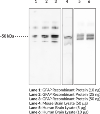
Glial fibrillary acidic protein (GFAP) is a protein encoded by the GFAP gene in humans and a member of the class III intermediate filament (IF) protein family.{53009} It is composed of an N-terminal head domain, a highly conserved α-helical rod domain, and a C-terminal tail domain that mediate GFAP self-assembly, dimerization, and oligomerization, respectively.{53010,53011} GFAP is expressed in, and has commonly been used as a pan marker for, mature astrocytes.{53009} GFAP IFs form a dynamic network of cytosolic filament proteins that collectively provide structure and strength to the cytoskeleton of astrocytes, thus supporting their morphology and function.{53009} Isolated astrocytes from neonatal Gfap-/- mouse brain have reduced numbers of IFs and IF bundles, increased proliferation, and loss of contact-inhibited growth.{53015,53016} Gfap-/- mice develop more diffuse and infiltrative brain lesions compared to wild-type littermates in a mouse model of experimental autoimmune encephalomyelitis (EAE).{53019} Mutations in the rod and tail domains of GFAP have been associated with Rosenthal fiber formation, a hallmark of Alexander disease.{53013} Transgenic overexpression of Gfap in mice increases the expression of certain cytokines and antioxidative enzymes in the olfactory bulb and has been used as a mouse model of Alexander disease.{53017} GFAP can be citrullinated on the arginine residue at position 270 (R270) and at R416 by protein arginine deiminase 1 (PAD1; Item No. 10784) and PAD2 (Item No. 10785).{53021} Citrullinated GFAP has been found in rat cerebral cortex in a model of traumatic brain injury, as well as in postmortem hippocampus from patients with Alzheimer’s disease.{53020,53021} Cayman’s GFAP Polyclonal Antibody can be used for ELISA, IHC, and WB applications. The antibody recognizes GFAP at ~50 kDa from human and murine samples.
Brand:CaymanSKU:28848 - 500 µgAvailable on backorder
 Brand:Boster BioSKU:EK1440
Available on backorder
Globotetraosylceramides (Item Nos. 24881) are bioactive neutral glycosphingolipids. They are the major glycolipids in human erythrocytes.{43016} They act as receptors for the Shiga toxins Stx1, Stx2, and Stx2e, the cytotoxic protein pierisin-1, and parvovirus B19.{43017,43018,43019} Globotetraosylceramides increase the expression of proteins responsible for enamel deposition, including ameloblastin, amelogenin, and enamelin, in dental epithelial cells and activate the ERK and p38 MAPK signaling pathways.{43020} Levels of globotetraosylceramides are elevated in fibroblasts from patients with salt and pepper syndrome, a neurocutaneous condition characterized by intellectual disability and hyper- and hypo-pigmented skin.{43021} Globotetraosylceramide Polyclonal Antibody can be used for ELISA and TLC immunoblotting. [Matreya, LLC. Catalog No. 1960]
Brand:CaymanSKU:28641 - 50 µlAvailable on backorder
Brand:CaymanSKU:600572 - 125 µlAvailable on backorder

Most histone lysine methyltransferases contain a conserved domain (SET) that utilizes S-adenosyl-L-methionine (SAM or AdoMet) as a co-factor to catalyze the methylation of the epsilon amino group of lysine. G9a-like protein (GLP) is a SET domain-containing methyltransferase that specifically mono- and di-methylates histone H3 at lysine 9 (H3K9). GLP and G9a function as major euchromatic H3K9me1 and H3K9me2 histone methyltransferases and also have been found to methylate several nonhistone substrates, including p53(K372). This fluorescence polarization assay is based upon a proprietary small molecule fluorescent probe* that binds to the SAM binding pocket in GLP. Binding of the small molecule probe to GLP induces an increase in fluorescence polarization. Binding of the probe can be competed with the endogenous cofactor SAM or by the inhibitor sinefungin, but is unaffected by the histone H3 peptide substrate. The GLP SAM-Screener™ Assay is robust (Z’ >0.5) and exhibits a greater than 100 mP shift over a range of 0-250 nM GLP. The assay is suitable for high-throughput screening in the provided 384-well plate or can be scaled to higher density plate formats (e.g., 1,536-well) if desired. *United States Patent 9,120,820
Brand:CaymanSKU:600570 - 1920 wellsAvailable on backorder
Most histone lysine methyltransferases contain a conserved domain (SET) that utilizes S-adenosyl-L-methionine (SAM or AdoMet) as a co-factor to catalyze the methylation of the epsilon amino group of lysine. G9a-like protein (GLP) is a SET domain-containing methyltransferase that specifically mono- and di-methylates histone H3 at lysine 9 (H3K9). GLP and G9a function as major euchromatic H3K9me1 and H3K9me2 histone methyltransferases and also have been found to methylate several nonhistone substrates, including p53(K372). This fluorescence polarization assay is based upon a proprietary small molecule fluorescent probe* that binds to the SAM binding pocket in GLP. Binding of the small molecule probe to GLP induces an increase in fluorescence polarization. Binding of the probe can be competed with the endogenous cofactor SAM or by the inhibitor sinefungin, but is unaffected by the histone H3 peptide substrate. The GLP SAM-Screener™ Assay is robust (Z’ >0.5) and exhibits a greater than 100 mP shift over a range of 0-250 nM GLP. The assay is suitable for high-throughput screening in the provided 384-well plate or can be scaled to higher density plate formats (e.g., 1,536-well) if desired. *United States Patent 9,120,820
Brand:CaymanSKU:600570 - 384 wellsAvailable on backorder
Brand:CaymanSKU:10010098 - 1 eaAvailable on backorder
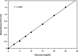
Glucose, a monosaccharide (or simple sugar), is the most important carbohydrate in biology. Transported via the blood stream, it is the primary source of energy for the body’s cells. Glucose level is tightly regulated in the human body. Failure to maintain blood glucose in the normal range leads to conditions of persistently high (hyperglycemia) or low (hypoglycemia) blood sugar. Diabetes mellitus, characterized by persistent hyperglycemia, is the most prominent disease related to failure of blood sugar regulation. Cayman’s Glucose Colorimetric Assay Kit provides a simple, reproducible, and sensitive tool for assaying glucose in plasma, serum, and urine. The glucose assay uses the glucose oxidase-peroxide reaction for the determination of glucose concentrations. In this assay, glucose is oxidized to δ-gluconolactone with concomitant reduction of the flavin adenine dinucleotide (FAD)-dependent enzyme glucose oxidase. The reduced form of glucose oxidase is regenerated to its oxidized form by molecular oxygen to produce hydrogen peroxide. Finally, with horseradish peroxidase as a catalyst, hydrogen peroxide reacts with 3,5-dichloro-2-hydroxybenzenesulfonic acid and 4-aminoantipyrine to generate a pink dye with an optimal absorption at 514 nm.
Brand:CaymanSKU:10009582 - 2 x 96 wellsAvailable on backorder
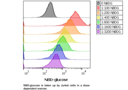
Glucose metabolism is a primary source of energy and biomaterials for the maintenance of cell homeostasis. Extra glucose is stored in the muscles and liver as glycogen which is hydrolyzed to glucose and released into the blood when needed. The rate of glucose uptake in cells is dynamic and tightly regulated by hormones and/or growth factors including insulin. Cancer cells exhibit increased glucose uptake and metabolism by aerobic glycolysis in order to support a high rate of proliferation. Chemicals that block glucose uptake by cancer cells have been shown to have anti-cancer effects. Cayman’s Glucose Uptake Cell-based Assay Kit provides a convenient tool for studying modulators of cellular glucose uptake. The kit employs 2-NBDG, a fluorescently labeled deoxyglucose analog, as a probe for the detection of glucose taken up by cultured cells. Apigenin, a flavonoid that has been reported to be an inhibitor of glucose transport, is included as a control.
Brand:CaymanSKU:600470 - 1 eaAvailable on backorder
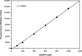
Glucose-6-phosphate dehydrogenase (G6PDH) is a cytosolic enzyme that catalyzes the first step in the pentose phosphate pathway. This pathway includes converting glucose to ribose-5-phosphate, a precursor to RNA, DNA, ATP, CoA, NAD, and FAD. The pathway also generates NADPH. G6PDH deficiency, the most common enzyme deficiency worldwide, causes a spectrum of diseases including neonatal hyperbilirubinemia, acute hemolysis, and chronic hemolysis. G6PDH activity has been shown to be upregulated in rat and mouse models of obesity, hyperglycemia, and hyperinsulinemia. Cayman’s Glucose-6-Phosphate Dehydrogenase Assay provides a fluorescence-based method for detecting G6PDH activity in a variety of samples including erythrocyte lysates, tissue homogenates, and cell culture samples.
Brand:CaymanSKU:700300 - 96 wellsAvailable on backorder
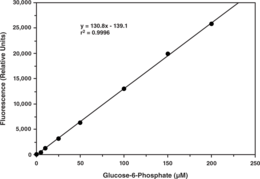
Glucose-6-phosphate (G6P) plays a key role in the homeostatic regulation of blood glucose levels by participating in glycolysis, glycogen synthesis, gluconeogenesis, and can be metabolized to NADPH, enabling protection against oxidative damage. Disruption of G6P activity leads to glycogen storage disease type I or von Gierke’s disease, a group of inherited metabolic diseases characterized by severe hypoglycemia, growth retardation, and hepatomegaly, due to accumulation of glycogen and fat in the liver. Cayman’s Glucose-6-Phosphate Fluorometric Assay provides a fluorescence-based method for detecting G6P in tissue homogenates and cell culture samples. In the assay, G6PDH catalyzes the oxidation of G6P to 6-phospho-D-gluconate, along with the concomitant reduction of NADP+ to NADPH. NADPH reacts with the fluorometric detector to yield a highly fluorescent product which can be analyzed with an excitation wavelength of 530-540 nm and an emission wavelength of 585-595 nm.
Brand:CaymanSKU:700750 - 96 wellsAvailable on backorder
The ion channels activated by glutamate are typically divided into two classes. Those that are sensitive to N-methyl-D-aspartate (NMDA) are designated NMDA receptors (NMDAR) while those activated by α-amino-3-hydroxy-5-methyl-4-isoxalone propionic acid (AMPA) are known as AMPA receptors (AMPAR). The AMPAR are comprised of four distinct glutamate receptor subunits designated (GluR1-4) and they play key roles in virtually all excitatory neurotransmission in the brain. The GluR1 subunit is widely expressed throughout the nervous system. GluR1 is potentiated by phosphorylation at Ser831 which has been shown to be mediated by either PKC or CaM kinase II. In addition, phosphorylation of this site has been linked to synaptic plasticity as well as learning and memory.
Brand:CaymanSKU:10602 - 1 eaAvailable on backorder
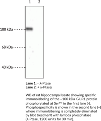
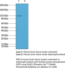
GluR1 is a subunit of the AMPA ionotropic glutamate receptor, which is responsible for fast excitatory synaptic transmission in the CNS.{59665} AMPA receptors are composed of four subunits, GluR1, GluR2, GluR3, and GluR4, which combine into heterotetramers to form a cation-permeable pore in the plasma membrane. Each subunit has two isoforms with the primary isoform designated as flip and a second isoform generated through alternative splicing designated as flop.{59667,52879} The GluR1 flip and flop isoforms do not affect desensitization or channel opening and closing kinetics.{52879} GluR1 can be phosphorylated by PKA at serine 845 (Ser845), which increases the peak open probability of the ion channel, and dephosphorylation is required for AMPA receptor endocytosis.{59665} GluR1 (phospho-Ser845) levels increase in the ventromedial prefrontal cortex (vmPFC) and nucleus accumbens (NAc) following cocaine-cue extinction.{59666} Levels of GluR1 (phospho-Ser845) are reduced following NMDA receptor activation in rat hippocampal slices and by amyloid-β (Aβ) oligomers in primary mouse neurons.{59668,59669} Cayman’s GluR1 (Phospho-Ser845) Polyclonal Antibody can be used for immunohistochemistry (IHC) and Western blot (WB) applications. The antibody recognizes Glur1 (phosphor-Ser845) at approximately 100 kDa from mouse, rat, and turtle samples.
Brand:CaymanSKU:10601 - 1 eaAvailable on backorder
GluR1 is a subunit of the AMPA ionotropic glutamate receptor, which is responsible for fast excitatory synaptic transmission in the CNS.{59665} AMPA receptors are composed of four subunits, GluR1, GluR2, GluR3, and GluR4, which combine into heterotetramers to form a cation-permeable pore in the plasma membrane. Each subunit has two isoforms with the primary isoform designated as flip and a second isoform generated through alternative splicing designated as flop.{59667,52879} The GluR1 flip and flop isoforms do not affect desensitization or channel opening and closing kinetics.{52879} GluR1 can be phosphorylated by PKA at serine 845 (Ser845), which increases the peak open probability of the ion channel, and dephosphorylation is required for AMPA receptor endocytosis.{59665} GluR1 (phospho-Ser845) levels increase in the ventromedial prefrontal cortex (vmPFC) and nucleus accumbens (NAc) following cocaine-cue extinction.{59666} Levels of GluR1 (phospho-Ser845) are reduced following NMDA receptor activation in rat hippocampal slices and by amyloid-β (Aβ) oligomers in primary mouse neurons.{59668,59669} Cayman’s GluR1 (Phospho-Ser845) Rabbit Monoclonal Antibody can be used for immunohistochemistry (IHC) and Western blot (WB) applications.
Brand:CaymanSKU:32242 - 100 µlAvailable on backorder
AMPA receptors are ionotropic glutamate receptors that mediate excitatory synaptic transmission.{46957,46955} They are tetrameric protein complexes expressed throughout the central nervous system in both neurons and glia that are assembled from combinations of GluR1, GluR2, GluR3, and GluR4, also known as GluR-A-D, subunits, each of which has extracellular N-terminal and ligand binding domains, a channel domain consisting of three membrane-spanning helices and a channel pore loop, and an intracellular C-terminus.{46955,46956,46958,46959} The GluR1 subunit, which is encoded by GRIA1 in humans, is primarily expressed in the forebrain and hippocampus. Mice lacking the GluR1 subunit exhibit normal spatial reference memory but have deficits in spatial working memory.{46957} They also exhibit increased and decreased duration of nocifensive behaviors in phases I and II, respectively, of the formalin test, as well as increased duration of nocifensive behaviors in response to intraplantar injection of capsaicin (Item No. 92350), compared with wild-type mice.{55099} SNPs in GRIA1 have been found in patients with bipolar disorder with psychotic features.{46958} Elevated levels of GRIA1 mRNA have been found in postmortem dorsolateral prefrontal cortex from patients with schizophrenia.{55100} Cayman’s GluR1 Monoclonal Antibody can be used for Western blot (WB) applications. The antibody recognizes GluR1 at approximately 105 kDa from mouse and rat samples.
Brand:CaymanSKU:29277 - 100 µlAvailable on backorder
AMPA receptors are ionotropic glutamate receptors that mediate excitatory synaptic transmission.{46955,46957} They are tetrameric protein complexes expressed throughout the CNS in both neurons and glia that are assembled from combinations of GluR1, GluR2, GluR3, and GluR4, also known as GluR-A-D, subunits, each of which has extracellular N-terminal and ligand-binding domains, a channel domain consisting of three membrane-spanning helices and a channel pore loop, and an intracellular C-terminus.{46955,46956,46958,46959} GluR1 and GluR2 are predominantly expressed in the forebrain with low levels of GluR3 and GluR4, and pyramidal neurons express AMPA receptors primarily comprised of heterotetramers of GluR1 and GluR2 subunits. GluR2 is encoded by GRIA2 and regulates AMPA receptor calcium permeability, assembly, trafficking, and single channel conductance.{46955,56145} It undergoes RNA editing at position 586, the Q/R site, to introduce an arginine residue that renders GluR2 subunit-containing AMPA receptors impermeable to calcium.{46955,56145,56146} Motor neurons isolated from Gria2-deficient mouse embryos exhibit increased calcium influx and increased susceptibility to AMPA receptor-mediated excitotoxicity.{56146} Heterozygous knockdown of Gria2 accelerates motor neuron degradation and decreases lifespan in the SOD1G93A transgenic mouse model of amyotrophic lateral sclerosis. GRIA2 expression is reduced in postmortem hippocampus from patients with chronic schizophrenia.{56147} Cayman’s GluR2 Polyclonal Antibody can be used for Western blot (WB) applications. The antibody detects GluR2 at approximately 100 kDa from rat samples.
Brand:CaymanSKU:29278 - 100 µlAvailable on backorder
AMPA receptors are ionotropic glutamate receptors that mediate excitatory synaptic transmission.{46955,46957} They are tetrameric protein complexes expressed throughout the central nervous system in both neurons and glia that are assembled from combinations of GluR1, GluR2, GluR3, and GluR4, also known as GluR-A-D, subunits, each of which has extracellular N-terminal and ligand binding domains, a channel domain consisting of three membrane-spanning helices and a channel pore loop, and an intracellular C-terminus.{46955,46956,46958,46959} GluR1 and GluR2 are predominantly expressed in the forebrain, with low levels of GluR3 and GluR4, and pyramidal neurons express AMPA receptors primarily comprised of heterotetramers of GluR1 and GluR2 subunits. Hippocampal pyramidal neurons also express AMPA receptors composed of GluR2 and GluR3 (GluR2/3) that cycle into and out of synapses constitutively.{46952} Protein levels of GluR2/3 are decreased in postmortem enterorhinal cortex, frontal cortex, and Purkinje cell samples from patients with sporadic Creutzfeldt-Jakob disease.{46953} GluR2/3 protein levels are increased on the dendrites of dentate granule cells in postmortem epileptogenic hippocampus samples from patients with temporal lobe epilepsy.{46954} Cayman’s GluR2/3 Polyclonal Antibody can be used for Western blot (WB) applications. The antibody recognizes GluR2/3 at approximately 100 kDa from rat samples.
Brand:CaymanSKU:29279 - 100 µlAvailable on backorder
Glutaredoxin 1 (Grx1) is a thiol-disulfide oxidoreductase and member of the thioredoxin family encoded by GLRX1 with a role in the maintenance of cellular thiol redox homeostasis.{53968} It is ubiquitously expressed and localizes to the cytosol, nucleus, and mitochondrial intermembrane space. Grx1 is a dithiol Grx that contains two active site cysteine residues, which catalyze the deglutathionylation of protein disulfides and glutathionylated proteins to regulate redox signal transduction and repair oxidized proteins.{49701,53969,49702} Knockdown or overexpression of GLRX1 enhances or reduces, respectively, oxidative stress-induced apoptosis in HEK293T cells.{49703} Glrx1-/- mice have increased bronchoalveolar lavage fluid (BALF) macrophage infiltration and lung glutathionylated protein levels induced by cigarette smoke compared with wild-type mice.{49704} Grx1 levels are decreased in postmortem brain from patients with Parkinson’s disease.{49702} Cayman’s Glutaredoxin 1 (human) Polyclonal Antibody can be used for immunohistochemistry (IHC) and Western blot (WB) applications. The antibody recognizes Grx1 from human samples.
Brand:CaymanSKU:11545 - 1 eaAvailable on backorder
Glutaredoxin 1 (Grx1) is a thiol-disulfide oxidoreductase and member of the thioredoxin family encoded by GLRX1 with a role in the maintenance of cellular thiol redox homeostasis.{53968} It is ubiquitously expressed and localizes to the cytosol, nucleus, and mitochondrial intermembrane space. Grx1 is a dithiol Grx that contains two active site cysteine residues, which catalyze the deglutathionylation of protein disulfides and glutathionylated proteins to regulate redox signal transduction and repair oxidized proteins.{49701,53969,49702} Knockdown or overexpression of GLRX1 enhances or reduces, respectively, oxidative stress-induced apoptosis in HEK293 T cells.{49703} Glrx1-/- mice have increased bronchoalveolar lavage fluid (BALF) macrophage infiltration and lung glutathionylated protein levels induced by cigarette smoke compared with wild-type mice.{49704} Grx1 levels are decreased in postmortem brain from patients with Parkinson’s disease.{49702} Cayman’s Glutaredoxin 1 (human) Polyclonal Antibody – Biotinylated can be used for ELISA, immunohistochemistry (IHC), and Western blot (WB) applications.
Brand:CaymanSKU:11546 - 1 eaAvailable on backorder
Glutaredoxin 2 (Grx2) is a thiol-disulfide oxidoreductase and member of the thioredoxin family encoded by GLRX2 with a role in the maintenance of cellular thiol redox homeostasis.{53968,53969} Alternative splicing of GLRX2 produces three ubiquitously expressed isoforms, Grx2a, Grx2b, and Grx2c, with Grx2a localized to the mitochondria and Grx2b and Grx2c in the cytoplasm and nucleus. It is a dithiol Grx that contains two active site cysteine residues and catalyzes the glutathione-dependent reduction of disulfides, acting as an electron donor for ribonucleotide or sulfate reduction, and regulating protein levels of glutathione mixed disulfides.{53969} In its inactive state, Grx2 is homodimer linked by a single [2Fe-2S] cluster, which acts as a redox sensor that drives monomerization and activation of Grx2 under conditions of oxidative stress, such as free radical formation or oxidation of the glutathione (GSH) pool.{53969,53970,53971} The oxidized active site in Grx2 is reduced by GSH and, unlike other eukaryotic Grxs, is also a substrate for thioredoxin reductase (TrxR). Grx2 catalyzes the reversible glutathionylation of mitochondrial complex I to regulate superoxide production and facilitates the reduction of protein disulfides, glutathionylated proteins, and other low molecular weight substrates under conditions of oxidative stress.{57138} Knockdown or overexpression of GLRX2 enhances and reduces, respectively, oxidative stress-induced apoptosis in HeLa cells. Glrx2 knockdown increases high-fat diet-induced insulin resistance, hippocampal inflammation, increases in body weight, and cognitive dysfunction in mice.{53972} Cayman’s Glutaredoxin 2 (human) Polyclonal Antibody can be used for Western blot (WB) applications.
Brand:CaymanSKU:11544 - 1 eaAvailable on backorder
Brand:CaymanSKU:11536 - 1 eaAvailable on backorder

Cayman’s GSH assay kit utilizes a carefully optimized enzymatic recycling method, using glutathione reductase for the quantification of GSH.{3644,3649,4674} The sulfhydryl group of GSH reacts with DTNB (5,5′-dithio-bis-2-nitrobenzoic acid, Ellman’s reagent) and produces a yellow colored 5-thio-2-nitrobenzoic acid (TNB). The mixed disulfide, GSTNB (between GSH and TNB) that is concomitantly produced, is reduced by glutathione reductase to recycle the GSH and produce more TNB. The rate of TNB production is directly proportional to this recycling reaction which in turn is directly proportional to the concentration of GSH in the sample. Measurement of the absorbance of TNB at 405 or 412 nm provides an accurate estimation of GSH in the sample. GSH is easily oxidized to the disulfide dimer GSSG. Because of the use of glutathione reductase in the Cayman GSH assay kit, both GSH and GSSG are measured and the assay reflects total glutathione. The kit can also be used to measure only GSSG by following an alternative protocol. GSH measurement can be done in plasma, tissue samples, and cultured cells using this kit. Nearly all samples require deproteination before assay.
Brand:CaymanSKU:703002 - 480 wellsAvailable on backorder
Cayman’s GSH assay kit utilizes a carefully optimized enzymatic recycling method, using glutathione reductase for the quantification of GSH.{3644,3649,4674} The sulfhydryl group of GSH reacts with DTNB (5,5′-dithio-bis-2-nitrobenzoic acid, Ellman’s reagent) and produces a yellow colored 5-thio-2-nitrobenzoic acid (TNB). The mixed disulfide, GSTNB (between GSH and TNB) that is concomitantly produced, is reduced by glutathione reductase to recycle the GSH and produce more TNB. The rate of TNB production is directly proportional to this recycling reaction which in turn is directly proportional to the concentration of GSH in the sample. Measurement of the absorbance of TNB at 405 or 412 nm provides an accurate estimation of GSH in the sample. GSH is easily oxidized to the disulfide dimer GSSG. Because of the use of glutathione reductase in the Cayman GSH assay kit, both GSH and GSSG are measured and the assay reflects total glutathione. The kit can also be used to measure only GSSG by following an alternative protocol. GSH measurement can be done in plasma, tissue samples, and cultured cells using this kit. Nearly all samples require deproteination before assay.
Brand:CaymanSKU:703002 - 96 wellsAvailable on backorder
Glutathione (GSH) is the most prevalent low molecular-weight tripeptide thiol in animal cells. It serves as a primary cellular anti-oxidant and plays a fundamental role in the elimination of environmental toxins. During early apoptosis, cells may exclude GSH, causing a decrease in intracellular GSH levels. The intracellular level of GSH is thus used as an indicator of cell health. Cayman’s Cell-Based Glutathione Assay Kit (Blue Fluorescence) employs a cell-permeable dye, monochlorobimane (MCB), which reacts with GSH to generate a highly fluorescent product that can be measured using excitation and emission wavelengths of 380 and 480 nm, respectively. The kit can be easily adapted to screening programs for therapeutic compounds regulating intracellular GSH levels.
Brand:CaymanSKU:600360 - 96 wellsAvailable on backorder
![The Glutathione Fluorescent Assay Kit is designed to measure the amount of total glutathione (GSH+GSSG) in cells and tissue homogenates and the total amount (including GSSG and protein mixed disulfides with glutathione) in plasma. The assay is simple and sensitive based on eosinGSH that emits a strong fluorescent signal detectable at 545 nm after excitation at 520 nm.[IMCO Catalog FkGSH-01]](https://interpriseusa.com/wp-content/uploads/2021/04/26404.png)
Glutathione (GSH) is a tripeptide (γ-Glu-Cys-Gly) present a very high concentrations throughout living organisms (from 1 to 10 nM) and exists in three major forms: reduced sulphydryl (GSH), glutathione disulfide (GSSG), or bound to Cys residues in proteins (PSSG). From its first described role in heavy metals and xenobiotics detoxification, it is now known that GSH plays a key role in cell redox homeostasis, gene expression, cell proliferation, and apoptosis mainly by binding to Cys residues in protein, known as protein glutathionylation (PSSG). GSH is used by glutaredoxin (Grx) to reduce disulfides. This reaction is usually coupled to the NADPH driven reduction of GSSG by glutathione reductase (GR). The ratio GSH/GSSG has been used as an indicator of the redox level in cells but this parameter can also be estimated by the quantification of PSSG. In fact, PSSG has the advantage of being more stable than GSSG. Within mammalian cells, glutathione exists mainly (>98%) in the thiol-reduced form (GSH). In fact the oxidized form, GSSG, is toxic in the cytosol and it is either reduced by glutathione reductase or exported from cells when its cytosolic concentration increases. Vice versa in plasma glutathione is present mainly as the oxidized form both as GSSG and mixed disulfides (Cysteinyl-glutathione and PSSG). This kit is designed to measure the amount of total glutathione (GSH+GSSG) in cells and tissue homogenates and the total amount (including GSSG and protein mixed disulfides with glutathione) in plasma.{35436} The assay is simple and sensitive based on eosinGSH that emits a strong fluorescent signal detectable at 545 nm after excitation at 520 nm.[IMCO Catalog FkGSH-01]
Brand:CaymanSKU:26404 - 96 wellsAvailable on backorder
Brand:CaymanSKU:703114 - 1 eaAvailable on backorder
Glutathione peroxidase (GPX) catalyzes the reduction of hydroperoxides, including hydrogen peroxides, by reduced glutathione and functions to protect the cell from oxidative damage. With the exception of phospholipid-hydroperoxide GPX, a monomer, all of the GPX enzymes are tetramers of four identical subunits.{7580,7585} Each subunit contains a selenocysteine in the active site which participates directly in the two-electron reduction of the peroxide substrate.{7580,7585} The enzyme uses glutathione as the ultimate electron donor to regenerate the reduced form of the selenocysteine.{7580,7585} The Cayman Chemical Glutathione Peroxidase Assay Kit measures GPX activity indirectly by a coupled reaction with glutathione reductase (GR). Oxidized glutathione (GSSG), produced upon reduction of an organic hydroperoxide by GPX, is recycled to its reduced state by GR and NADPH. The oxidation of NADPH to NADP+ is accompanied by a decrease in absorbance at 340 nm. The rate of decrease in the A340 is directly proportional to the GPX activity in the sample.{7579} The Cayman GPX Assay Kit can be used to measure all of the glutathione-dependent peroxidases in plasma, erythrocyte lysates, tissue homogenates, and cell lysates.
Brand:CaymanSKU:703102 - 480 wellsAvailable on backorder
Glutathione peroxidase (GPX) catalyzes the reduction of hydroperoxides, including hydrogen peroxides, by reduced glutathione and functions to protect the cell from oxidative damage. With the exception of phospholipid-hydroperoxide GPX, a monomer, all of the GPX enzymes are tetramers of four identical subunits.{7580,7585} Each subunit contains a selenocysteine in the active site which participates directly in the two-electron reduction of the peroxide substrate.{7580,7585} The enzyme uses glutathione as the ultimate electron donor to regenerate the reduced form of the selenocysteine.{7580,7585} The Cayman Chemical Glutathione Peroxidase Assay Kit measures GPX activity indirectly by a coupled reaction with glutathione reductase (GR). Oxidized glutathione (GSSG), produced upon reduction of an organic hydroperoxide by GPX, is recycled to its reduced state by GR and NADPH. The oxidation of NADPH to NADP+ is accompanied by a decrease in absorbance at 340 nm. The rate of decrease in the A340 is directly proportional to the GPX activity in the sample.{7579} The Cayman GPX Assay Kit can be used to measure all of the glutathione-dependent peroxidases in plasma, erythrocyte lysates, tissue homogenates, and cell lysates.
Brand:CaymanSKU:703102 - 96 wellsAvailable on backorder
Glutathione reductase (GR, EC 1.6.4.2) is a flavoprotein that catalyzes the NADPH-dependent reduction of oxidized glutathione (GSSG) to glutathione (GSH).{7626} This enzyme is essential for the GSH redox cycle which maintains adequate levels of reduced cellular GSH. A high GSH/GSSG ratio is essential for protection against oxidative stress. The Cayman Chemical Glutathione Reductase Assay Kit measures GR activity by measuring the rate of NADPH oxidation. The oxidation of NADPH to NADP+ is accompanied by a decrease in absorbance at 340 nm. Since GR is present at rate limiting concentrations, the rate of decrease in the A340 is directly proportional to the GR activity in the sample. The Cayman GR Assay Kit is provided in a convenient 96 well plate format and can be used to measure GR activity in plasma, erythrocyte lysates, tissue homogenates, and cell lysates.
Brand:CaymanSKU:703202 - 96 wellsAvailable on backorder

Glutathione S-transferases (GSTs) are ubiquitous multifunctional enzymes, which play a key role in cellular detoxification. The enzymes protect cells against toxicants by conjugating them to glutathione, thereby neutralizing their electrophilic sites, and rendering the products more water-soluble.{8335} The glutathione conjugates are metabolized further to mercapturic acid and then excreted.{8335} Based on their sequence homology, substrate specificity, and immunological cross-reactivity, GSTs have been grouped into species-independent classes of isozymes.{8202,7934,8203,8387} These classes are comprised of cytosolic enzymes and the fifth form is microsomal.{8202,7934,8203,8387} The Cayman Chemical Glutathione S-Transferase Assay Kit measures total GST activity (cytosolic and microsomal) by measuring the conjugation of 1-chloro-2,4-dinitrobenzene (CDNB) with reduced glutathione.{7935} The conjugation is accompanied by an increase in absorbance at 340 nm. The rate of increase is directly proportional to the GST activity in the sample. The Cayman GST Assay Kit can be used to measure GST activity in plasma, erythrocyte lysates, tissue homogenates, and cell lysates. Cytosolic and microsomal GST activity can also be assayed separately.
Brand:CaymanSKU:703302 - 96 wellsAvailable on backorder
Cayman Chemical’s Glutathione S-Transferase (GST) Polyclonal Antibody may be used to monitor expression of recombinant proteins tagged with GST (GST fusion proteins). A recombinant GST-tagged protein is expected to migrate 26 kDa slower than the non-tagged version in reducing SDS-PAGE. In general, GSTs are ubiquitous multifunctional enzymes, which play a key role in cellular detoxification, and researchers routinely utilize the enzyme to simplify the detection or purification of recombinant proteins.{8335}
Brand:CaymanSKU:10010013 - 1 eaAvailable on backorder
Cayman’s Glutathione S-Transferase (GST) Polyclonal FITC Antibody may be used to monitor espression of recombinant proteins tagged with GST (GST fusion proteins). A recombinant GST-tagged protein is expected to migrate 26 kDa slower than the non-tagged version in reducing conditions on SDS-PAGE. In general, GSTs are ubiquitous multifunctional enzymes, which play a key role in cellular detoxification. Researchers routinely utilize the enzyme as a method to simplify the purification of recombinant proteins.{8335}
Brand:CaymanSKU:10197 - 500 µlAvailable on backorder

In mammals, triglycerides are constantly synthesized from fatty acids and segregated into cytosolic lipid droplets, mainly in adipocytes, as the major energy storage depot. During fasting, triglycerides stored in adipose tissue and liver are hydrolyzed by hormone-sensitive lipase and adipose triglyceride lipase to produce free fatty acids and glycerol. Cayman’s Cell-Based Assay Kit provides a convenient tool for studying triglycerides/fatty acid cycling and its regulation in adipocytes or hepatocytes. This kit will allow investigators to screen compounds involved in lipid storage and metabolism. Chloroquine is included in the kit as a positive control for screening pharmaceuticals that induce lipid droplet accumulation and free glycerol release from hepatocytes.
Brand:CaymanSKU:10011725 - 1 eaAvailable on backorder

Glycerol is the backbone of triglycerides and an important intermediate in energy metabolism being involved in both oxidative and synthetic processes. The measurement of circulating levels of glycerol and free fatty acids are considered to reflect lipolysis and are therefore useful parameters to evaluate in various conditions. The measurements of glycerol is also useful for correction of glycerol interference when measuring triglycerides in reference materials and in serum from patients with elevated glycerol concentrations. Cayman’s Glycerol Assay provides a simple, reproducible, and sensitive tool for measurement of glycerol in plasma and serum. The assay employs a coupled enzymatic reaction system that yields a brilliant purple product with an absorbance maximum at 540 nm.
Brand:CaymanSKU:10010755 - 96 wellsAvailable on backorder
Glycine is an important inhibitory transmitter in the brain stem and spinal cord. Glycine receptors are members of the ligand-gated ion channel family (LGICs) that mediate rapid chemical neurotransmission.{14450} The binding of glycine to its receptor produces a large increase in chloride conductance, which causes membrane hyperpolarization. Glycine receptors are anchored at inhibitory chemical synapses by a cytoplasmic protein, gephyrin.{14448} The glycine receptor has been used to great advantage in the identification of the binding sites for alcohol on the LGIC family of proteins.{14445,14449} These receptors have also been extremely useful in studies of synaptic clustering of receptors.{14447} The glycine receptor may also act in concert with an NMDAR subunit to form an excitatory receptor.{14446}
Brand:CaymanSKU:10009399 - 1 eaAvailable on backorder
Brand:CaymanSKU:700482 - 14 mlAvailable on backorder
Glycogen is a polysaccharide that is the principal storage form of glucose in animal and human cells. Glycogen is made primarily by the liver and muscles, but it can also be made by glycogenesis within the brain and stomach. Cayman’s Glycogen Assay provides a simple, reproducible, and sensitive tool for assaying glycogen from tissue. In this assay, glycogen is hydrolyzed by amyloglucosidase to form β-D-glucose, which is then specifically oxidized to D-glucono-δ-lactone by glucose oxidase forming hydrogen peroxide in the process. Hydrogen peroxide, in the presence of horseradish peroxidase, reacts with 10-acetyl-3,7-dihydroxyphenoxazine (ADHP) in a 1:1 stoichiometry to generate the highly fluorescent product resorufin which is measure at an excitation wavelength of 530-540 nm and an emission wavelength of 585-595 nm.
Brand:CaymanSKU:700480 - 96 wellsAvailable on backorder
Brand:CaymanSKU:600454 - 250 µlAvailable on backorder
Brand:CaymanSKU:600452 - 1 eaAvailable on backorder
Brand:CaymanSKU:600451 - 250 µlAvailable on backorder

Cayman’s Glycolysis Cell-Based Assay Kit provides a colorimetric method for detecting L-lactate, the end product of glycolysis, produced and secreted by cultured cells. In this assay, lactate dehydrogenase catalyzes the reaction between NAD+ and lactate, yielding pyruvate and NADH. The NADH directly reduces a tetrazolium salt (INT) to a colored formazan which absorbs between 490 and 520 nm. The quantity of formazan produced is proportional to the quantity of lactate in the culture medium, and is thus an indirect measurement of glycolysis. This assay can be adapted to high-throughput screening to examine glycolytic regulators in a cell-based system. In addition, the non-invasive sampling leaves the cells intact and thus allows multiplex testing for additional markers.
Brand:CaymanSKU:600450 - 1 eaAvailable on backorder
Brand:CaymanSKU:10008856 - 200 µlAvailable on backorder
Goat anti-Chicken IgY conjugated to SureLight® APC Antibody: Goat polyclonal anti-chicken IgY Dye: SureLight® Allophycocyanin (APC) Excitation max. λ: 650 nm Emission max. λ: 660 nm Uses: Flow cytometry and cell-based assays
Brand:CaymanSKU:13391 - 100 μgAvailable on backorder
Goat anti-Chicken IgY conjugated to SureLight® R-PE Antibody: Goat polyclonal anti-chicken IgY Dye: SureLight® R-phycoerythrin Excitation max. λ: 565>498 nm Emission max. λ: 578 nm Uses: Flow cytometry, cell-based assays, microarrays, and microplate applications
Brand:CaymanSKU:13413 - 1 eaAvailable on backorder
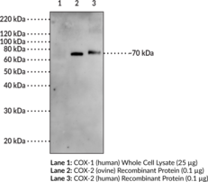
Cyclooxygenase (COX) catalyzes the first step in the biosynthesis of prostaglandins (PGs), thromboxanes, and prostacyclins: the conversion of arachidonic acid to PGH2. Discoveries of the induction of COX by a variety of stimuli such as phorbol esters, lipopolysaccharides, and cytokines led to the hypothesis that the inducible form of COX, COX-2, is responsible for the biosynthesis of PGs under acute inflammatory conditions.{4,3463} COX-2 is a 72 kDa protein which has been cloned from a variety of species including human, mouse, rat, and ovine.{2,7,2884,4051}
Brand:CaymanSKU:100034 - 1 eaAvailable on backorder
Goat anti-GST (Glutathione-S-Transferase) IgG conjugated to Alkaline Phosphatase Antibody: Goat polyclonal anti-Glutathione-S-Transferase Enzyme: Alkaline Phosphatase Uses: Western blot detection. Recommended dilution of 1:500-1:10,000. Starting dilution: 1:2,000.
Brand:CaymanSKU:13461 - 100 μgAvailable on backorder
Goat anti-GST (Glutathione-S-Transferase) IgG conjugated to DyLight® 488 Antibody: Goat polyclonal anti-GST (Glutathione-S-Transferase) IgG Dye: DyLight® 488 Excitation max. λ: 493 nm Emission max. λ: 518 nm Uses: Flow cytometry, cell-based assays, microarrays, and microplate applications
Brand:CaymanSKU:11245 - 100 μgAvailable on backorder
Goat anti-GST (Glutathione-S-Transferase) IgG conjugated to DyLight® 550 Antibody: Goat polyclonal anti-GST (Glutathione-S-Transferase) IgG Dye: DyLight® 550 Excitation max. λ: 562 nm Emission max. λ: 576 nm Uses: Flow cytometry, cell-based assays, microarrays, and microplate applications
Brand:CaymanSKU:11254 - 100 μgAvailable on backorder
Goat anti-GST (Glutathione-S-Transferase) IgG conjugated to DyLight® 650 Antibody: Goat polyclonal anti-GST (Glutathione-S-Transferase) IgG Dye: DyLight® 650 Excitation max. λ: 652 nm Emission max. λ: 672 nm Uses: Flow cytometry, cell-based assays, microarrays, and microplate applications
Brand:CaymanSKU:11263 - 100 μgAvailable on backorder
Antibody: Goat polyclonal anti-GST (Glutathione-S-Transferase) IgG Dye: Europium Chelate W-1024 Excitation max. λ: 340 nm Emission max. λ: 615 nm Uses: TR-FRET high throughput screening (HTS) applications (LANCE®, HTRF®)
Brand:CaymanSKU:16575 - 1 mgAvailable on backorder
Antibody: Goat polyclonal anti-GST (Glutathione-S-Transferase) IgG Dye: Europium Chelate W-1024 Excitation max. λ: 340 nm Emission max. λ: 615 nm Uses: TR-FRET high throughput screening (HTS) applications (LANCE®, HTRF®)
Brand:CaymanSKU:16575 - 20 µgAvailable on backorder
Antibody: Goat polyclonal anti-GST (Glutathione-S-Transferase) IgG Dye: Europium Chelate W-1024 Excitation max. λ: 340 nm Emission max. λ: 615 nm Uses: TR-FRET high throughput screening (HTS) applications (LANCE®, HTRF®)
Brand:CaymanSKU:16575 - 50 µgAvailable on backorder
Goat anti-GST (Glutathione-S-Transferase) IgG conjugated to SureLight® APC Antibody: Goat polyclonal anti-GST (Glutathione-S-Transferase) IgG Dye: SureLight® Allophycocyanin (APC) Excitation max. λ: 652 nm Emission max. λ: 657.5 nm Uses: Optimized for High throughput screening (HTS) and fluorescence resonance energy transfer (FRET) assays, flow cytometry, and microscopy
Brand:CaymanSKU:16598 - 1 mgAvailable on backorder
Goat anti-GST (Glutathione-S-Transferase) IgG conjugated to SureLight® APC Antibody: Goat polyclonal anti-GST (Glutathione-S-Transferase) IgG Dye: SureLight® Allophycocyanin (APC) Excitation max. λ: 652 nm Emission max. λ: 657.5 nm Uses: Optimized for High throughput screening (HTS) and fluorescence resonance energy transfer (FRET) assays, flow cytometry, and microscopy
Brand:CaymanSKU:16598 - 100 µgAvailable on backorder
Goat anti-GST (Glutathione-S-Transferase) IgG conjugated to SureLight® P3 Antibody: Goat polyclonal anti-Glutathione-S-Transferase (GST) Dye: SureLight® P3 Excitation max. λ: 614 nm Emission max. λ: 662 nm Uses: Flow cytometry, cell-based assays, microarrays, and microplate applications
Brand:CaymanSKU:13448 - 100 μgAvailable on backorder
Goat anti-GST (Glutathione-S-Transferase) IgG conjugated to SureLight® PerCP Antibody: Goat polyclonal anti-GST (Glutathione-S-Transferase) IgG Dye: SureLight® Peridinin-Chlorophyll (PerCP) Excitation max. λ: 483 nm Emission max. λ: 673 nm Uses: Flow cytometry, cell-based assays, microarrays, and microplate applications
Brand:CaymanSKU:11272 - 100 μgAvailable on backorder
Goat anti-GST (Glutathione-S-Transferase) IgG conjugated to SureLight® R-PE Antibody: Goat polyclonal anti-GST (Glutathione-S-Transferase) IgG Dye: SureLight® R-phycoerythrin Excitation max. λ: 565>498 nm Emission max. λ: 578 nm Uses: Flow cytometry, cell-based assays, microarrays, and microplate applications
Brand:CaymanSKU:13422 - 1 eaAvailable on backorder
Goat anti-Human IgG, H+L conjugated to HRP Antibody: Goat polyclonal anti-Human IgG Enzyme: Horseradish Peroxidase Uses: Western blot detection. Recommended dilution of 1:500-1:10,000. Starting dilution: 1:2,000.
Brand:CaymanSKU:16703 - 1 mgAvailable on backorder
Spectral Characteristics: Visible absorption maximum = 652 nm, Emission maximum = 657 nm; This polyclonal antibody detects human IgA, tagged with SureLight® APC.
Brand:CaymanSKU:16584 - 100 µgAvailable on backorder
Goat anti-Human IgA conjugated to SureLight® R-PE Antibody: Goat polyclonal anti-Human IgA Dye: SureLight® R-phycoerythrin Excitation max. λ: 565>498 nm Emission max. λ: 578 nm Uses: Flow cytometry, cell-based assays, microarrays, and microplate applications
Brand:CaymanSKU:16639 - 100 µgAvailable on backorder
Goat anti-Human IgE conjugated to SureLight® APC Antibody: Goat polyclonal anti-Human IgE Dye: SureLight® Allophycocyanin (APC) Excitation max. λ: 650 nm Emission max. λ: 660 nm Uses: Flow cytometry and cell-based assays
Brand:CaymanSKU:16585 - 100 µgAvailable on backorder