ELISA Kits
Showing 3451–3600 of 3623 results
-
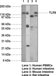
The toll-like receptors (TLRs) in mammals comprise a family of transmembrane proteins characterized by multiple copies of leucine rich repeats in the extracellular domain and an interleukin-1 (IL-1) receptor motif in the cytoplasmic domain. Like their counterparts in Drosophila, TLRs signal through adaptor molecules.{17463} The TLR family is a phylogenetically conserved mediator of innate immunity that is essential for microbial recognition.{17464} Most mammalian species have between ten and fifteen types of TLRs. Ten functional TLRs (TLR1-10) have been identified in human. Humans also encode a TLR11 gene but it contains several stop codons and protein is not expressed. However, mouse and rat TLR11 are functional, and it is thought that human TLR11 function was lost during evolution. Historically speaking, TLR expression has been most extensively studied in the immune system. Overall, TLRs are highly expressed in immune competent cells, including macrophages, dendritic cells, neutrophils, mucosal epithelial cells and dermal endothelial cells. However, TLRs have also been identified in many other cell types and anatomical tissue locations where they are expressed either constitutively or induced during infection. Gene knockout experiments suggest that TLR9 acts as a receptor for unmethylated CpG dinucleotides in bacterial DNA.{17576} Human and mouse TLR9 share an overall amino acid identity of 75.5%. TLR9 is highly expressed in spleen.
Brand:CaymanSKU:13593 - 1 eaAvailable on backorder
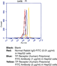
Thromboxane A2 (TXA2) is a potent vasoconstrictor and activator of platelet aggregation. TXA2 elicits its effects via a 7-transmembrane domain G protein-coupled receptor, the TP receptor.{3168} The TP receptor is highly expressed in platelets and is relatively less abundant in tissues such as lung, kidney, brain, spleen, thymus, monocytes, uterus, and placenta.{3167,11368,2061,4311} The apparent molecular weight for TP receptors has been reported from 37 kDa to 70 kDa, depending on different degrees of glycosylation.{8380,4311,7235} However Cayman’s antibody consistently detects the TP receptor at 55 and 64 kDa in platelet, kidney, and Cos-7 cell samples. Cayman Chemical’s TP receptor polyclonal antibody can be used for western blot and immunocytochemical analysis for both the TPA and TPB splice variants of the receptor on samples of human, murine, rat, and COS-7 (African green monkey) origin.
Brand:CaymanSKU:10004452 - 1 eaAvailable on backorder
Thromboxane A2 (TXA2) is a potent vasoconstrictor and activator of platelet aggregation. The short half-life of TXA2 ensures local action whether generated by vascular endothelial cells or by platelets and confers physiologically beneficial or deleterious effects under inflammatory situations.{146,11131} TXA2 elicits its effects via a 7-transmembrane domain G protein-coupled receptor, the TP receptor.{6062} This receptor can also bind prostaglandin H2 and isoprostanes and was first cloned from human placenta and the platelet-like MEG-01 cell line.{11146,7729} The TP receptor is highly expressed in platelets and is relatively less abundant in tissues such as lung, kidney, brain, spleen, thymus, monocytes, uterus, and placenta.{3167,11368,2061,4311,8380,7235}
Brand:CaymanSKU:10012559 - 1 eaAvailable on backorder

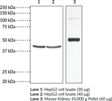
Thromboxane A2 (TXA2) is a potent vasoconstrictor and activator of platelet aggregation. TXA2 elicits its effects via a 7-transmembrane domain G-protein coupled receptor, the TP receptor.{6062} The TP receptor is highly expressed in platelets and is relatively less abundant in tissues such as lung, kidney, brain, spleen, thymus, monocytes, uterus, and placenta.{3167,11368,2061,4311} The apparent molecular weight for TP receptors has been reported from 37 kDa to 70 kDa, depending on different degrees of glycosylation.{8380,4311,7235} However, Cayman’s antibody consistently detects the TP receptor at 42 kDa in kidney, Hep2, and COS-7 cell samples. Cayman’s TP receptor polyclonal antibody can be used for Western blot analysis for both the TPA and TPB splice variants of the receptor on samples of human, mouse, rat, monkey and bovine origin. Use of this antibody for other species and applications has not yet been tested.
Brand:CaymanSKU:101882 - 1 eaAvailable on backorder
Brand:CaymanSKU:600503 - 10 mlAvailable on backorder
Brand:CaymanSKU:600503 - 2 mlAvailable on backorder
Brand:CaymanSKU:600508 - 10 mlAvailable on backorder
Brand:CaymanSKU:600508 - 2 mlAvailable on backorder
Brand:CaymanSKU:600504 - 1 gAvailable on backorder
Brand:CaymanSKU:600504 - 200 mgAvailable on backorder
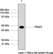
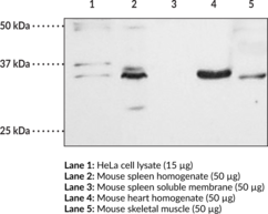
Tumor necrosis factor (TNF)-induced signaling is mediated through association of TNF receptor (TNFR) with adaptor proteins, such as TNF receptor-associated factors (TRAFs). TRAFs form a family of cytoplasmic adapter proteins that mediate signal transduction from many members of the TNF-receptor superfamily (e.g., RANK, CD30, CD40, etc.) and the interleukin-1 receptor. The carboxy-terminal region of TRAFs is required for self-association and interaction with receptor cytoplasmic domains following ligand-induced oligomerization. Recent molecular cloning studies have led to identification of seven TRAFs (TRAF1-TRAF7).{19431,18761,19383,19384,19385} Human TRAF5 is a 557-amino acid protein. TRAF5 is implicated in NF-kB and c-Jun NH(2)-terminal kinase/stress-activated protein kinase activation by members of the TNF receptor superfamily, including CD27, CD30, CD40, and lymphotoxin-β receptor. Targeted disruption of TRAF5 gene causes defects in CD40-CD27 mediated lymphocyte activation.{19385}
Brand:CaymanSKU:10873 - 1 eaAvailable on backorder
TNF-related apoptosis-inducing ligand (TRAIL) is a protein encoded by the TNFSF10 gene in humans and is a member of the TNF superfamily of proteins.{52585} TRAIL contains an N-terminal cytoplasmic domain, a transmembrane domain, and a C-terminal extracellular receptor binding domain.{52585,52586} It is produced in immune effector cells as a transmembrane precursor protein from which mature, soluble TRAIL is formed by proteolysis.{52586} Soluble TRAIL is cleaved from the precursor protein on the extracellular side of the membrane in response to stimulation by cytokines, such as IFN-γ (Item No. 32008).{52585} TRAIL monomers form trimers with a single zinc atom bound at the trimer interface, which is required for its structural stability and function.{52587} The trimer binds to the pro-apoptotic death receptors DR4 and DR5 and the decoy receptors DcR1 and DcR2, which lack functional intracellular domains.{52585} The intracellular death domains of DR4 or DR5 colocalize upon TRAIL binding, which recruits Fas-associated death domain (FADD) and pro-caspase-8 and initiates either the extrinsic or intrinsic apoptotic pathways depending on the cell type. DR4 and DR5 are highly expressed on a variety of cancer cells while DcR1 and DcR1 are primarily expressed on non-cancerous cells, which allows TRAIL to selectively induce apoptosis in cancer cells.{52588} Recombinant human TRAIL induces apoptosis in a variety of cancer cell lines and reduces tumor growth in mouse xenograft models.{52589} However, it increases proliferation in certain cancer cells in vitro, and other cells lines develop resistance, which can sometimes be partially mitigated by combining it with other agents. A cysteine-to-threonine mutation at position 723 of TNFSF10 is associated with sporadic breast cancer, and SNPS in TNFSF10 are associated with fatty liver disease, multiple sclerosis, and asthma.{52585} TNFSF10 expression is decreased in breast cancer patients with brain metastases and increased in patients with multiple sclerosis. In addition, protein levels of TRAIL are increased in patients with systemic lupus erythematous (SLE) and multiple sclerosis. Cayman’s TRAIL Polyclonal Antibody can be used for Western blot (WB) applications. The antibody recognizes TRAIL at approximately 35 kDa from human samples.
Brand:CaymanSKU:160750 - 500 µlAvailable on backorder
Brand:CaymanSKU:10006882 - 1 eaAvailable on backorder
Brand:CaymanSKU:10006880 - 1 eaAvailable on backorder
Brand:CaymanSKU:10008895 - 1 eaAvailable on backorder
Brand:CaymanSKU:10008897 - 1 eaAvailable on backorder
Brand:CaymanSKU:10008896 - 1 eaAvailable on backorder
Brand:CaymanSKU:10009279 - 1 eaAvailable on backorder
Brand:CaymanSKU:10006884 - 1 eaAvailable on backorder
Brand:CaymanSKU:10009269 - 1 eaAvailable on backorder
Brand:CaymanSKU:10007883 - 1 eaAvailable on backorder
Brand:CaymanSKU:600592 - 120 µlAvailable on backorder
Brand:CaymanSKU:601082 - 120 µlAvailable on backorder
Brand:CaymanSKU:600023 - 1 eaAvailable on backorder
Brand:CaymanSKU:10007442 - 1 eaAvailable on backorder
Brand:CaymanSKU:10006883 - 1 eaAvailable on backorder
Brand:CaymanSKU:10007444 - 1 eaAvailable on backorder
Brand:CaymanSKU:10010894 - 1 eaAvailable on backorder
Brand:CaymanSKU:10008859 - 1 eaAvailable on backorder
Transcription Factor STAT3 Primary Antibody has been tested and formulated to work exclusively with Cayman’s STAT3 Transcription Factor Assay Kit (Item No. 601950). Please visit STAT3 Transcription Factor Assay Kit (Item No. 601950) for the kit protocol, procedures, and product handling.
Brand:CaymanSKU:601951 - 1 eaAvailable on backorder
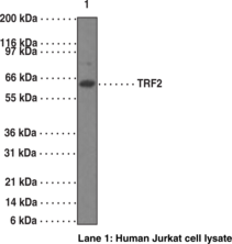
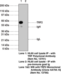
Telomeric repeat binding factor (TRF2) is a ubiquitously expressed protein that is implicated in the control of telomere length.{18142} TRF2, like TRF1, contains a Myb-related DNA binding motif. It binds to duplex TTAGGG repeats and is localized to all human telomeres in metaphase chromosomes.{18142} TRF2 is thought to protect chromosome ends by maintaining the correct structure at telomere termini.{18143} The use of mutant forms of TRF2 has implicated a role of TRF2 in the prevention of senescence in primary human cells.{18143} Inhibition of TRF2 results in apoptosis in a subset of mammalian cell types.{18144}
Brand:CaymanSKU:13790 - 1 eaAvailable on backorder
Telomeric repeat binding factor (TRF2) is a ubiquitously expressed protein that is implicated in the control of telomere length.{18142} TRF2, like TRF1, contains a Myb-related DNA binding motif. It binds to duplex TTAGGG repeats and is localized to all human telomeres in metaphase chromosomes.{18142} TRF2 is thought to protect chromosome ends by maintaining the correct structure at telomere termini.{18143} The use of mutant forms of TRF2 has implicated a role of TRF2 in the prevention of senescence in primary human cells.{18143} Inhibition of TRF2 results in apoptosis in a subset of mammalian cell types.{18144}
Brand:CaymanSKU:13791 - 1 eaAvailable on backorder
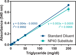
The measurement of triglyceride levels, in conjunction with other lipid assays, are useful in the diagnosis of primary and secondary hyperlipoproteinemia, dyslipidemia, and triglyceridemia. Cayman’s Triglyceride Assay Kit provides a simple, reproducible, and sensitive tool for assaying triglycerides in plasma, serum, cell lysates, and tissue homogenate samples. The assay is initiated with the enzymatic hydrolysis of the triglycerides by lipase to produce glycerol and free fatty acids. The glycerol released is subsequently measured by a coupled enzymatic reaction system with a colorimetric readout at 540 nm.
Brand:CaymanSKU:10010303 - 96 wellsAvailable on backorder
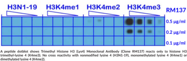
Histone H3 undergoes many modifications including acetylation, methylation, and phosphorylation that are important for regulation of gene transcription. Methylation at lysine 4 of histone 3 (H3K4) is an evolutionarily conserved modification associated with transcriptionally active chromatin. The SET1/MLL (KMT2) family of methyltransferases is the principal enzyme family responsible for H3K4 methylations. This antibody reacts to histone H3 trimethylated at lysine 4 (K4me3) and does not detect monomethylated lysine 4 (K4me1), dimethylated lysine 4 (K4me2), or other methylations in histone H3.
Brand:CaymanSKU:20718 - 100 µgAvailable on backorder
Brand:CaymanSKU:700751 - 5 mlAvailable on backorder
Ion channels are integral membrane proteins that help establish and control the small voltage gradient across the plasma membrane of living cells by allowing the flow of ions down their electrochemical gradient.{17533} They are present in the membranes that surround all biological cells and their main function is to regulate the flow of ions across this membrane. Whereas some ion channels permit the passage of ions based on charge, others conduct based on a ionic species, such as sodium or potassium. Furthermore, in some ion channels, the passage is governed by a gate which is controlled by chemical or electrical signals, temperature, or mechanical forces. There are a few main classifications of gated ion channels. There are voltage-gated ion channels, ligand-gated, other gating systems, and finally those that are classified differently, having more exotic characteristics. The first are voltage-gated ion channels which open and close in response to membrane potential. These are then seperated into sodium, calcium, potassium, proton, transient receptor, and cyclic nucleotide-gated channels, each of which is responsible for a unique role. Ligand-gated ion channels are also known as ionotropic receptors and they open in response to specific ligand molecules binding to the extracellular domain of the receptor protein. The other gated classifications include activation and inactivation by second messengers, inward-rectifier potassium channels, calcium-activated potassium channels, two-pore-domain potassium channels, light-gated channels, mechano-sensitive ion channels, and cyclic nucleotide-gated channels. Finally, the other classifications are based on less normal characteristics such as two-pore channels and transient receptor potential channels.{17535} Transient receptor potential cation channel, subfamily C, member 4 (TRPC4), is a human gene encoding a protein of the same name. They are expressed in smooth muscle and endothelial cells where they regulate membrane potential and calcium influx. TRPC4 is activated by G(q)/phospholipase C-coupled receptors, but the underlying mechanism remains elusive.{17697} Studies suggest TRPC4 contributes to axonal regeneration after nerve injury.{17698}
Brand:CaymanSKU:13719 - 100 µgAvailable on backorder
Ion channels are integral membrane proteins that help establish and control the small voltage gradient across the plasma membrane of living cells by allowing the flow of ions down their electrochemical gradient.{17533} They are present in the membranes that surround all biological cells and their main function is to regulate the flow of ions across this membrane. Whereas some ion channels permit the passage of ions based on charge, others conduct based on a ionic species, such as sodium or potassium. Furthermore, in some ion channels, the passage is governed by a gate which is controlled by chemical or electrical signals, temperature, or mechanical forces. There are a few main classifications of gated ion channels. There are voltage-gated ion channels, ligand-gated, other gating systems, and finally those that are classified differently, having more exotic characteristics. The first are voltage-gated ion channels which open and close in response to membrane potential. These are then seperated into sodium, calcium, potassium, proton, transient receptor, and cyclic nucleotide-gated channels, each of which is responsible for a unique role. Ligand-gated ion channels are also known as ionotropic receptors and they open in response to specific ligand molecules binding to the extracellular domain of the receptor protein. The other gated classifications include activation and inactivation by second messengers, inward-rectifier potassium channels, calcium-activated potassium channels, two-pore-domain potassium channels, light-gated channels, mechano-sensitive ion channels, and cyclic nucleotide-gated channels. Finally, the other classifications are based on less normal characteristics such as two-pore channels and transient receptor potential channels.{17535} TRPs, mammalian homologs of the Drosophila transient receptor potential (TRP) protein, are ion channels that are thought to mediate capacitative calcium entry into the cell. TRP-PLIK is a protein that is both an ion channel and a kinase. As a channel, it conducts calcium and monovalent calcium. As a kinase, it is capable of phosphorylating itself and other substrates. The kinase activity is necessary for channel function, as shown by its dependence on intracellular ATP and by the kinase mutant.{17703,17704}
Brand:CaymanSKU:13720 - 100 µgAvailable on backorder
Ion channels are integral membrane proteins that help establish and control the small voltage gradient across the plasma membrane of living cells by allowing the flow of ions down their electrochemical gradient.{17533} They are present in the membranes that surround all biological cells and their main function is to regulate the flow of ions across this membrane. Whereas some ion channels permit the passage of ions based on charge, others conduct based on a ionic species, such as sodium or potassium. Furthermore, in some ion channels, the passage is governed by a gate which is controlled by chemical or electrical signals, temperature, or mechanical forces. There are a few main classifications of gated ion channels. There are voltage-gated ion channels, ligand-gated, other gating systems, and finally those that are classified differently, having more exotic characteristics. The first are voltage-gated ion channels which open and close in response to membrane potential. These are then seperated into sodium, calcium, potassium, proton, transient receptor, and cyclic nucleotide-gated channels, each of which is responsible for a unique role. Ligand-gated ion channels are also known as ionotropic receptors and they open in response to specific ligand molecules binding to the extracellular domain of the receptor protein. The other gated classifications include activation and inactivation by second messengers, inward-rectifier potassium channels, calcium-activated potassium channels, two-pore-domain potassium channels, light-gated channels, mechano-sensitive ion channels, and cyclic nucleotide-gated channels. Finally, the other classifications are based on less normal characteristics such as two-pore channels and transient receptor potential channels.{17535} The TRPV3 proteins belongs to a family of nonselective cation channels that function in a variety of processes, including temperature sensation and vasoregulation. The thermosensitive members of this family are expressed in subsets of sensory neurons that terminate in the skin and are activated at distinct physiological temperatures. This channel is activated at temperatures between 22 and 40°C. The gene lies in close proximity to another family member (TRPV1) gene on chromosome 17, and the two encoded proteins are thought to associate with each other to form heteromeric channels.{17705,17706}
Brand:CaymanSKU:13721 - 100 µgAvailable on backorder
 Brand:CaymanSKU:32864 - 100 µl
Available on backorder
 Brand:CaymanSKU:32865 - 100 µl
Available on backorder
Brand:CaymanSKU:10009092 - 1 eaAvailable on backorder
TPH catalyzes the 5-hydroxylation of tryptophan, which is the first step in the biosynthesis of indoleamines (serotonin and melatonin).{14424} In mammals, serotonin biosynthesis occurs predominantly in neurons which originate in the Raphe nuclei of the brain, and melatonin synthesis takes place within the pineal gland. TPH catalyzes the 5-hydroxylation of tryptophan, which is the first step in the biosynthesis of indoleamines (serotonin and melatonin).{14424} In mammals, serotonin biosynthesis occurs predominantly in neurons which originate in the Raphe nuclei of the brain, and melatonin synthesis takes place within the pineal gland. Although TPH catalyzes the same reaction within the Raphe nuclei and the pineal gland, TPH activity is rate-limiting for serotonin but not melatonin biosynthesis. Serotonin functions mainly as a neurotransmitter, whereas melatonin is the principal hormone secreted by the pineal gland. The activity of TPH is enhanced by phosphorylation by cAMP-dependent protein kinase (PKA) and Ca2+/calmodulin kinase II (CaM K II).{14425,14426} CaM K II phosphorylate Ser260 which lies within the regulatory domain of TPH.{14425}
Brand:CaymanSKU:10009398 - 1 eaAvailable on backorder
Tryptophan hydroxylase (TPH) catalyzes the 5-hydroxylation of tryptophan, which is the first step in the biosynthesis of indoleamines (serotonin and melatonin).{14424} In mammals, serotonin biosynthesis occurs predominantly in neurons which originate in the Raphe nuclei of the brain, and melatonin synthesis takes place within the pineal gland. Although TPH catalyzes the same reaction within the Raphe nuclei and the pineal gland, TPH activity is rate-limiting for serotonin but not melatonin biosynthesis. Serotonin functions mainly as a neurotransmitter, whereas melatonin is the principal hormone secreted by the pineal gland. The activity of TPH is enhanced by phosphorylation by cAMP-dependent protein kinase (PKA) and Ca2+/calmodulin kinase II (CaM K II).{14425,14426} Both PKA and CaM K II phosphorylate Ser58 which lies within the regulatory domain of TPH.{14427}
Brand:CaymanSKU:10009397 - 1 eaAvailable on backorder
Tryptophan hydroxylase (TPH) catalyzes the first step in the biosynthesis of serotonin and melatonin.{14424} Thus, expression of TPH can be used as an indicator of the localization of serotonin and melatonin in brain. In mammals, serotonin biosynthesis occurs predominantly in neurons which originate in the Raphe nuclei of the brain, and melatonin synthesis takes place within the pineal gland.{14423} Although TPH catalyzes the same reaction within the Raphe nuclei and the pineal gland, TPH activity is rate-limiting for serotonin but not melatonin biosynthesis.{14424}
Brand:CaymanSKU:10009396 - 1 eaAvailable on backorder
Tyrosine hydroxylase (TH) is the rate-limiting enzyme in the synthesis of the catecholamines dopamine and norepinephrine. TH antibodies can therefore be used as markers for dopaminergic and noradrenergic neurons in a variety of applications including depression, schizophrenia, Parkinson’s disease, and drug abuse.{14414,14416,14417} TH antibodies can also be used to explore basic mechanisms of dopamine and norepinephrine signalling.{14444,14443,14439} The activity of TH is also regulated by phosphorylation.{14441,14440,14442} Phospho-specific antibodies for the phosphorylation sites on TH can be used to great effect in studying this regulation and in identifying the cells in which TH phosphorylation occurs.
Brand:CaymanSKU:10009413 - 1 eaAvailable on backorder
Tyrosine hydroxylase (TH) is the rate-limiting enzyme in the synthesis of the catecholamines dopamine and norepinephrine. TH antibodies can therefore be used as markers for dopaminergic and noradrenergic neurons in a variety of applications including depression, schizophrenia, Parkinson’s disease, and drug abuse.{14414,14416,14417} TH antibodies can also be used to explore basic mechanisms of dopamine and norepinephrine signaling.{14444,14443,14439} The activity of TH is also regulated by phosphorylation.{14440,14441,14442} Phospho-specific antibodies for the phosphorylation sites on TH can be used to great effect in studying this regulation and in identifying the cells in which TH phosphorylation occurs.
Brand:CaymanSKU:10009414 - 1 eaAvailable on backorder
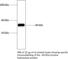
Tyrosine hydroxylase (TH) catalyzes the conversion of tyrosine to L-DOPA, which is the rate-limiting step in the biosynthesis of the catecholamines dopamine, norepinephrine, and epinephrine.{56214} It assembles into tetramers, with each monomer comprised of an N-terminal regulatory domain with three serine residues at positions 19, 31, and 40 that are subject to regulatory phosphorylation, a central catalytic domain, and a C-terminal tetramerization domain. TH is expressed in dopaminergic neurons in the olfactory bulb, diencephalon, substantia nigra, ventral tegmental area, and retinal amacrine cells, adrenergic and noradrenergic cells in the hypothalamus, medulla, and locus coeruleus (LC), as well as sympathetic ganglia and adrenal chromaffin cells. Due to its expression in catecholaminergic neurons, TH has been used as a marker of these cells in the brain.{56215} Mutations in TH have been found in patients with tyrosine hydroxylase deficiency, infantile parkinsonism, and progressive infantile encephalopathy.{56216,56217} Cayman’s Tyrosine Hydroxylase (rat, denatured) Polyclonal Antibody can be used for immunocytochemistry (ICC), immunohistochemistry (IHC), and Western Blot (WB) applications. The antibody recognizes TH at approximately 60 kDa from mammalian samples.
Brand:CaymanSKU:10604 - 1 eaAvailable on backorder
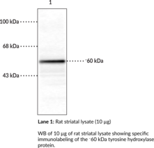
Tyrosine hydroxylase (TH) catalyzes the conversion of tyrosine to L-DOPA, which is the rate-limiting step in the biosynthesis of the catecholamines dopamine, norepinephrine, and epinephrine.{56214} It assembles into tetramers, with each monomer comprised of an N-terminal regulatory domain with three serine residues at positions 19, 31, and 40 that are subject to regulatory phosphorylation, a central catalytic domain, and a C-terminal tetramerization domain. TH is expressed in dopaminergic neurons in the olfactory bulb, diencephalon, substantia nigra, ventral tegmental area, and retinal amacrine cells, adrenergic and noradrenergic cells in the hypothalamus, medulla, and locus coeruleus (LC), as well as sympathetic ganglia and adrenal chromaffin cells. Due to its expression in catecholaminergic neurons, TH has been used as a marker of these cells in the brain.{56215} Mutations in TH have been found in patients with tyrosine hydroxylase deficiency, infantile parkinsonism, and progressive infantile encephalopathy.{56216,56217} Cayman’s Tyrosine Hydroxylase (rat, native) Polyclonal Antibody can be used for immunocytochemistry (ICC), immunohistochemistry (IHC), and Western blot (WB) applications. The antibody recognizes TH at approximately 60 kDa from human, mouse, rabbit, and rat samples.
Brand:CaymanSKU:29299 - 100 µlAvailable on backorder
Brand:CaymanSKU:700018 - 100 µlAvailable on backorder
Ubiquitin is a small protein that occurs in all eukaryotic cells. The ubiquitin protein itself consists of 76 amino acids and has a molecular mass of about 8.5 kDa. Key features include its C-terminal tail and the 7 Lys residues. It is highly conserved among eukaryotic species: human and yeast ubiquitin share 96% sequence identity.{17707} The main function of ubiquitin is to clear abnormal, foreign and improperly folded proteins by targeting them for degradation by the 26S proteasome.{17708} Ubiquitination represents an essential cellular process affected by a multi-enzyme cascade involving classes of enzymes known as ubiquitin-activating enzymes (E1s), ubiquitin-conjugating enzymes (E2s or Ubcs) and ubiquitin-protein ligases (E3s). Ubiquitin is activated in a two-step reaction by an E1 ubiquitin-activating enzyme in a process requiring ATP as an energy source. The initial step involves production of a ubiquitin-adenylate intermediate. The second step transfers ubiquitin to the E1 active site cysteine residue, with release of AMP. This step results in a thioester linkage between the C-terminal carboxyl group of ubiquitin and the E1 cysteine sulfhydryl group. The third step is a transfer of ubiquitin from E1 to the active site cysteine of a ubiquitin-conjugating enzyme E2 via a trans(thio)esterification reaction. And the final step of the ubiquitylation cascade creates an isopeptide bond between a lysine of the target protein and the C-terminal glycine of ubiquitin. In general, this step requires the activity of one of the hundreds of E3 ubiquitin-protein ligases (often termed simply ubiquitin ligase). E3 enzymes function as the substrate recognition modules of the system and are capable of interaction with both E2 and substrate.{17708,17709} Ubiquitination also participates in the internalization and degradation of plasma membrane proteins such as some of the TCR subunits while still ER-membrane associated.{17712} Ubiquitin also plays a role in regulating signal transduction cascades through the elimination inhibitor proteins, such as IκBα and p27.{17713}
Brand:CaymanSKU:13722 - 200 µgAvailable on backorder
Ubiquitin is a small protein that occurs in all eukaryotic cells. The ubiquitin protein itself consists of 76 amino acids and has a molecular mass of about 8.5 kDa. Key features include its C-terminal tail and the 7 Lys residues. It is highly conserved among eukaryotic species: human and yeast ubiquitin share 96% sequence identity.{17707} The main function of ubiquitin is to clear abnormal, foreign and improperly folded proteins by targeting them for degradation by the 26S proteasome.{17708} Ubiquitination represents an essential cellular process affected by a multi-enzyme cascade involving classes of enzymes known as ubiquitin-activating enzymes (E1s), ubiquitin-conjugating enzymes (E2s or Ubcs) and ubiquitin-protein ligases (E3s). Ubiquitin is activated in a two-step reaction by an E1 ubiquitin-activating enzyme in a process requiring ATP as an energy source. The initial step involves production of a ubiquitin-adenylate intermediate. The second step transfers ubiquitin to the E1 active site cysteine residue, with release of AMP. This step results in a thioester linkage between the C-terminal carboxyl group of ubiquitin and the E1 cysteine sulfhydryl group. The third step is a transfer of ubiquitin from E1 to the active site cysteine of a ubiquitin-conjugating enzyme E2 via a trans(thio)esterification reaction. And the final step of the ubiquitylation cascade creates an isopeptide bond between a lysine of the target protein and the C-terminal glycine of ubiquitin. In general, this step requires the activity of one of the hundreds of E3 ubiquitin-protein ligases (often termed simply ubiquitin ligase). E3 enzymes function as the substrate recognition modules of the system and are capable of interaction with both E2 and substrate.{17708,17709} Ubiquitination also participates in the internalization and degradation of plasma membrane proteins such as some of the TCR subunits while still ER-membrane associated.{17712} Ubiquitin also plays a role in regulating signal transduction cascades through the elimination inhibitor proteins, such as IκBα and p27.{17713}
Brand:CaymanSKU:13722 - 50 µgAvailable on backorder
Ubiquitin is a small protein that occurs in all eukaryotic cells. The ubiquitin protein itself consists of 76 amino acids and has a molecular mass of about 8.5 kDa. Key features include its C-terminal tail and the 7 Lys residues. It is highly conserved among eukaryotic species: human and yeast ubiquitin share 96% sequence identity.{17707} The main function of ubiquitin is to clear abnormal, foreign and improperly folded proteins by targeting them for degradation by the 26S proteasome.{17708} Ubiquitination represents an essential cellular process affected by a multi-enzyme cascade involving classes of enzymes known as ubiquitin-activating enzymes (E1s), ubiquitin-conjugating enzymes (E2s or Ubcs) and ubiquitin-protein ligases (E3s). Ubiquitin is activated in a two-step reaction by an E1 ubiquitin-activating enzyme in a process requiring ATP as an energy source. The initial step involves production of a ubiquitin-adenylate intermediate. The second step transfers ubiquitin to the E1 active site cysteine residue, with release of AMP. This step results in a thioester linkage between the C-terminal carboxyl group of ubiquitin and the E1 cysteine sulfhydryl group. The third step is a transfer of ubiquitin from E1 to the active site cysteine of a ubiquitin-conjugating enzyme E2 via a trans(thio)esterification reaction. And the final step of the ubiquitylation cascade creates an isopeptide bond between a lysine of the target protein and the C-terminal glycine of ubiquitin. In general, this step requires the activity of one of the hundreds of E3 ubiquitin-protein ligases (often termed simply ubiquitin ligase). E3 enzymes function as the substrate recognition modules of the system and are capable of interaction with both E2 and substrate.{17708,17709} Ubiquitination also participates in the internalization and degradation of plasma membrane proteins such as some of the TCR subunits while still ER-membrane associated.{17712} Ubiquitin also plays a role in regulating signal transduction cascades through the elimination inhibitor proteins, such as IκBα and p27.{17713}
Brand:CaymanSKU:13723 - 200 µgAvailable on backorder
Ubiquitin is a small protein that occurs in all eukaryotic cells. The ubiquitin protein itself consists of 76 amino acids and has a molecular mass of about 8.5 kDa. Key features include its C-terminal tail and the 7 Lys residues. It is highly conserved among eukaryotic species: human and yeast ubiquitin share 96% sequence identity.{17707} The main function of ubiquitin is to clear abnormal, foreign and improperly folded proteins by targeting them for degradation by the 26S proteasome.{17708} Ubiquitination represents an essential cellular process affected by a multi-enzyme cascade involving classes of enzymes known as ubiquitin-activating enzymes (E1s), ubiquitin-conjugating enzymes (E2s or Ubcs) and ubiquitin-protein ligases (E3s). Ubiquitin is activated in a two-step reaction by an E1 ubiquitin-activating enzyme in a process requiring ATP as an energy source. The initial step involves production of a ubiquitin-adenylate intermediate. The second step transfers ubiquitin to the E1 active site cysteine residue, with release of AMP. This step results in a thioester linkage between the C-terminal carboxyl group of ubiquitin and the E1 cysteine sulfhydryl group. The third step is a transfer of ubiquitin from E1 to the active site cysteine of a ubiquitin-conjugating enzyme E2 via a trans(thio)esterification reaction. And the final step of the ubiquitylation cascade creates an isopeptide bond between a lysine of the target protein and the C-terminal glycine of ubiquitin. In general, this step requires the activity of one of the hundreds of E3 ubiquitin-protein ligases (often termed simply ubiquitin ligase). E3 enzymes function as the substrate recognition modules of the system and are capable of interaction with both E2 and substrate.{17708,17709} Ubiquitination also participates in the internalization and degradation of plasma membrane proteins such as some of the TCR subunits while still ER-membrane associated.{17712} Ubiquitin also plays a role in regulating signal transduction cascades through the elimination inhibitor proteins, such as IκBα and p27.{17713}
Brand:CaymanSKU:13723 - 50 µgAvailable on backorder
Ubiquitin is a small protein that occurs in all eukaryotic cells. The ubiquitin protein itself consists of 76 amino acids and has a molecular mass of about 8.5 kDa. Key features include its C-terminal tail and the 7 Lys residues. It is highly conserved among eukaryotic species: human and yeast ubiquitin share 96% sequence identity.{17707} The main function of ubiquitin is to clear abnormal, foreign and improperly folded proteins by targeting them for degradation by the 26S proteasome.{17708} Ubiquitination represents an essential cellular process affected by a multi-enzyme cascade involving classes of enzymes known as ubiquitin-activating enzymes (E1s), ubiquitin-conjugating enzymes (E2s or Ubcs) and ubiquitin-protein ligases (E3s). Ubiquitin is activated in a two-step reaction by an E1 ubiquitin-activating enzyme in a process requiring ATP as an energy source. The initial step involves production of a ubiquitin-adenylate intermediate. The second step transfers ubiquitin to the E1 active site cysteine residue, with release of AMP. This step results in a thioester linkage between the C-terminal carboxyl group of ubiquitin and the E1 cysteine sulfhydryl group. The third step is a transfer of ubiquitin from E1 to the active site cysteine of a ubiquitin-conjugating enzyme E2 via a trans(thio)esterification reaction. And the final step of the ubiquitylation cascade creates an isopeptide bond between a lysine of the target protein and the C-terminal glycine of ubiquitin. In general, this step requires the activity of one of the hundreds of E3 ubiquitin-protein ligases (often termed simply ubiquitin ligase). E3 enzymes function as the substrate recognition modules of the system and are capable of interaction with both E2 and substrate.{17708,17709} Ubiquitination also participates in the internalization and degradation of plasma membrane proteins such as some of the TCR subunits while still ER-membrane associated.{17712} Ubiquitin also plays a role in regulating signal transduction cascades through the elimination inhibitor proteins, such as IκBα and p27.{17713}
Brand:CaymanSKU:13724 - 200 µgAvailable on backorder
Ubiquitin is a small protein that occurs in all eukaryotic cells. The ubiquitin protein itself consists of 76 amino acids and has a molecular mass of about 8.5 kDa. Key features include its C-terminal tail and the 7 Lys residues. It is highly conserved among eukaryotic species: human and yeast ubiquitin share 96% sequence identity.{17707} The main function of ubiquitin is to clear abnormal, foreign and improperly folded proteins by targeting them for degradation by the 26S proteasome.{17708} Ubiquitination represents an essential cellular process affected by a multi-enzyme cascade involving classes of enzymes known as ubiquitin-activating enzymes (E1s), ubiquitin-conjugating enzymes (E2s or Ubcs) and ubiquitin-protein ligases (E3s). Ubiquitin is activated in a two-step reaction by an E1 ubiquitin-activating enzyme in a process requiring ATP as an energy source. The initial step involves production of a ubiquitin-adenylate intermediate. The second step transfers ubiquitin to the E1 active site cysteine residue, with release of AMP. This step results in a thioester linkage between the C-terminal carboxyl group of ubiquitin and the E1 cysteine sulfhydryl group. The third step is a transfer of ubiquitin from E1 to the active site cysteine of a ubiquitin-conjugating enzyme E2 via a trans(thio)esterification reaction. And the final step of the ubiquitylation cascade creates an isopeptide bond between a lysine of the target protein and the C-terminal glycine of ubiquitin. In general, this step requires the activity of one of the hundreds of E3 ubiquitin-protein ligases (often termed simply ubiquitin ligase). E3 enzymes function as the substrate recognition modules of the system and are capable of interaction with both E2 and substrate.{17708,17709} Ubiquitination also participates in the internalization and degradation of plasma membrane proteins such as some of the TCR subunits while still ER-membrane associated.{17712} Ubiquitin also plays a role in regulating signal transduction cascades through the elimination inhibitor proteins, such as IκBα and p27.{17713}
Brand:CaymanSKU:13724 - 50 µgAvailable on backorder
This EIA so called Easy Sampling EIA kit works with any sample collected on any kind of protease inhibitors, without extraction but a simple dilution. Ghrelin discovered in 1999, is fast becoming an endocrinology target of the millennium. Ghrelin, identified in rat stomach as an endogenous ligand for the GH secretagogue receptor, is mainly produced in stomach, but has been demonstrated in many otther organs. In addition to GH-releasing properties and its orexant action, Ghrelin could act as an hormone having effects on gastric motility (similarity with the peptide hormone motilin), acidic secretion, cardiovascular action, antiproliferative effects, pancreatic and glucose metabolism function, sleep… Ghrelin gene raises to mRNA prepro-ghrelin of 117 amino acids. This precursor is processed into Ghrelin, 28 amino acids (human). Before being secreted, this peptide is octanoylated at Ser 3 by GOAT (Ghrelin Octanoyl Acyl Transferase). This step is essential for biological activity making GOAT a perfect target for drugs in feeding behaviour. Interestingly, the potential therapeutic importance of this hormone is not restricted to regulation of food intake but also in cachexia (related to cancer treatment, anorexia nervosa or ischemia) gastric motility and may be involved in osteoporosis, somatopause, infertility and ovulation induction, neurological disorders (Alcoholism, Post Traumatic Stress disorders…) and cardiovascular diseases. [Bertin Catalog No. A05320.96 wells]
Brand:CaymanSKU:501200 - 96 wellsAvailable on backorder
This EIA so called Easy Sampling EIA kit works with any sample collected on any kind of protease inhibitors, without extraction but a simple dilution. Ghrelin discovered in 1999, is fast becoming an endocrinology target of the millennium. Ghrelin, identified in rat stomach as an endogenous ligand for the GH secretagogue receptor, is mainly produced in stomach, but has been demonstrated in many other organs. In addition to GH-releasing properties and its orexant action, Ghrelin could act as an hormone having effects on gastric motility (similarity with the peptide hormone motilin), acidic secretion, cardiovascular action, antiproliferative effects, pancreatic and glucose metabolism function, sleep… Ghrelin gene raises to mRNA prepro-ghrelin of 117 amino acids. This precursor is processed into Ghrelin, 28 amino acids (human). Before being secreted, this peptide is octanoylated at Ser 3 by GOAT (Ghrelin Octanoyl Acyl Transferase). This step is essential for biological activity making GOAT a perfect target for drugs in feeding behaviour. Interestingly, the potential therapeutic importance of this hormone is not restricted to regulation of food intake but also in cachexia (related to cancer treatment, anorexia nervosa or ischemia) gastric motility and may be involved in osteoporosis, somatopause, infertility and ovulation induction, neurological disorders (Alcoholism, Post Traumatic Stress disorders…) and cardiovascular diseases. [Bertin Catalog No. A05319.96 wells]
Brand:CaymanSKU:501190 - 96 wellsAvailable on backorder
Ghrelin, an endogenous ligand for the growth hormone secretagogue receptor, is synthesized principally in the stomach. It stimulates food intake and transduces signals to hypothalamic regulatory nucleid that controls energy homeostasis. The peptide consists of 28 amino acids, with a n-octanoylation of the serine-3 residue, which is necessary for biological activity. Ghrelin is present in the peripheral circulation in two forms: acylated and unacylated. This EIA kit specifically measures the unacylated form of ghrelin. Each kit contains ghrelin (unacylated) AChE-Fab’ conjugate, human unacylated ghrelin standard, human unacylated ghrelin quality control, Ellman’s reagent, EIA buffer, wash buffer, Tween 20, plates pre-coated with anti-ghrelin mouse monoclonal antibody, and complete instructions. [Bertin Catalog No. A05119]
Brand:CaymanSKU:10008952 - 96 wellsAvailable on backorder
This EIA so called Easy Sampling EIA kit works with any sample collected on any kind of protease inhibitors, without extraction but a simple dilution. Ghrelin discovered in 1999, is fast becoming an endocrinology target of the millennium. Ghrelin, identified in rat stomach as an endogenous ligand for the GH secretagogue receptor, is mainly produced in stomach, but has been demonstrated in many other organs. In addition to GH-releasing properties and its orexant action, Ghrelin could act as an hormone having effects on gastric motility (similarity with the peptide hormone motilin), acidic secretion, cardiovascular action, antiproliferative effects, pancreatic and glucose metabolism function, sleep… Ghrelin gene raises to mRNA prepro-ghrelin of 117 amino acids. This precursor is processed into Ghrelin, 28 amino acids (human). Before being secreted, this peptide is octanoylated at Ser 3 by GOAT (Ghrelin Octanoyl Acyl Transferase). This step is essential for biological activity making GOAT a perfect target for drugs in feeding behaviour. Interestingly, the potential therapeutic importance of this hormone is not restricted to regulation of food intake but also in cachexia (related to cancer treatment, anorexia nervosa or ischemia) gastric motility and may be involved in osteoporosis, somatopause, infertility and ovulation induction, neurological disorders (Alcoholism, Post Traumatic Stress disorders…) and cardiovascular diseases. [Bertin Catalog No. A05318.96 wells]
Brand:CaymanSKU:501180 - 96 wellsAvailable on backorder
Ghrelin, an endogenous ligand for the growth hormone secretagogue receptor, is synthesized principally in the stomach. It stimulates food intake and transduces signals to hypothalamic regulatory nuclei that control energy homeostasis. The peptide consists of 28 amino acids, with an octanoylation of the serine-3 residue, which is necessary for biological activity. Ghrelin is present in the peripheral circulation in two forms: acylated and unacylated. This EIA kit specifically measures the unacylated form of ghrelin. Each kit contains ghrelin (unacylated) AChE-Fab’ conjugate, rat unacylated ghrelin standard, rat unacylated ghrelin quality control, Ellman’s reagent, EIA buffer, wash buffer, Tween 20, plates pre-coated with anti-ghrelin mouse monoclonal antibody, and complete instructions. [Bertin Catalog No. A05118]
Brand:CaymanSKU:10008953 - 96 wellsAvailable on backorder
Ghrelin discovered in 1999, is fast becoming an endocrinology target of the millennium. Ghrelin, identified in rat stomach as an endogenous ligand for the GH secretagogue receptor, is mainly produced in stomach, but has been demonstrated in many otther organs. In addition to GH-releasing properties and its orexant action, Ghrelin could act as an hormone having effects on gastric motility (similarity with the peptide hormone motilin), acidic secretion, cardiovascular action, antiproliferative effects, pancreatic and glucose metabolism function, sleep… Ghrelin gene raises to mRNA prepro-ghrelin of 117 amino acids. This precursor is processed into Ghrelin, 28 amino acids (human). Before being secreted, this peptide is octanoylated at Ser 3 by GOAT (Ghrelin Octanoyl Acyl Transferase). This step is essential for biological activity making GOAT a perfect target for drugs in feeding behaviour. Interestingly, the potential therapeutic importance of this hormone is not restricted to regulation of food intake but also in cachexia (related to cancer treatment, anorexia nervosa or ischemia) gastric motility and may be involved in osteoporosis, somatopause, infertility and ovulation induction, neurological disorders (Alcoholism, Post Traumatic Stress disorders…) and cardiovascular diseases. [Bertin Catalog No. A05402.96 wells]
Brand:CaymanSKU:501230 - 96 wellsAvailable on backorder
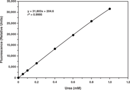
Urea or carbamide is synthesized in the body of many organisms as part of the urea cycle. It is the final degradation product of protein and amino acid metabolism. Urea is found in blood and is excreted by the kidney as a component of urine. Besides its role as carrier of waste nitrogen, urea also plays a role in the countercurrent exchange system of the nephrons, allowing for re-absorption of water and critical ions from the excreted urine. Cayman’s Urea Assay provides a convenient method for detecting urea in plasma, serum, and urine. In this assay, urease catalyzes the hydrolysis of urea into carbon dioxide and ammonia. Ammonia reacts with the detector resulting in a fluorescent product. Fluorescence is analyzed with an excitation wavelength of 405-415 nm and an emission wavelength of 470-480 nm.
Brand:CaymanSKU:700620 - 96 wellsAvailable on backorder
Brand:CaymanSKU:700622 - 1 eaAvailable on backorder
Brand:CaymanSKU:700321 - 5 mlAvailable on backorder

Uric acid (urate) is the end product of human purine metabolism and is mainly excreted in urine. Many factors, including genetic components and acquired factors, such as obesity and alcohol consumption, influence serum uric acid concentrations. Hyperuricemia induces or facilitates gout, kidney stones, metabolic syndrome, hypertension, and renal and cardiovascular disease, while exercise-induced acute renal failure is a significant complication of renal hypouricemia. Cayman’s Uric Acid Assay provides a fluorescence-based method for detecting uric acid in serum, plasma, and urine.
Brand:CaymanSKU:700320 - 96 wellsAvailable on backorder
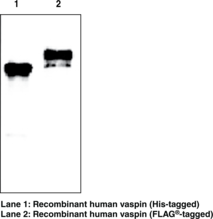
Vaspin, a serine protease inhibitor (serpin), is an insulin-sensitizing adipocytokine that has been isolated from both visceral and subcutaneous white adipose tissue. Based on recent findings, vaspin is suggested to regulate immune responses and inflammation and was found to be correlated with various metabolic parameters. Vaspin may represent a novel biomarker for obesity and impaired insulin sensitivity and might serve as a new therapeutic target of metabolic syndrome. Vaspin (human) monoclonal antibody (clone VP63) recognizes human vaspin. It detects a band of ~48 kDa by western blot.
Brand:CaymanSKU:10812 - 100 µgAvailable on backorder
Vaspin, a serine protease inhibitor (serpin), is an insulin-sensitizing adipocytokine that has been isolated from both visceral and subcutaneous white adipose tissue. Based on recent findings, vaspin is suggested to regulate immune responses and inflammation and was found to be correlated with various metabolic parameters. Vaspin may represent a novel biomarker for obesity and impaired insulin sensitivity and might serve as a new therapeutic target of metabolic syndrome. Vaspin (human) monoclonal antibody (clone VP63) recognizes human vaspin. It detects a band of ~48 kDa by western blot.
Brand:CaymanSKU:10812 - 50 µgAvailable on backorder
Cadherins are calcium-dependent cell adhesion proteins. They preferentially interact with themselves in a homophilic manner in connecting cells; cadherins may thus contribute to the sorting of heterogeneous cell types. VE-cadherin may play a important role in endothelial cell biology through control of the cohesion and organization of the intercellular junctions. It associates with alpha-catenin forming a link to the cytoskeleton. It acts in concert with KRIT1 to establish and maintain correct endothelial cell polarity and vascular lumen. [Bertin Catalog No. A03144]
Brand:CaymanSKU:32754 - 100 µgAvailable on backorder
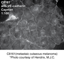
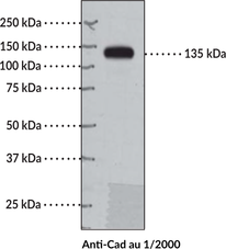
VE-cadherin (vascular endothelium cadherin), also known as Cadherin-5, is an endothelial cell-specific adhesion molecule localized at intracellular junctions.{7201, 7068,7071} The protein belongs to the cadherin superfamily of adhesion molecules.{7072} VE-cadherin plays a pivotal role in vascular structure assembly. Endothelial cells fail to organize in a vessel-like pattern if the VE-cadherin gene is disrupted.{7069} In this aspect, VE-cadherin is a critical component of a protein complex which controls endothelial cell tube formation.{7070} VE-cadherin is also critical for maintenance of the endothelium. Following disruption of endothelial cell monolayers with an anti-VE-cadherin antibody, endothelial cells rapidly resynthesize VE-cadherin in order to restore their intracellular junctions.{6997} Human VE-cadherin has a molecular mass of approximately 87,500 based on the deduced amino acid sequence and is 83% and 79% homologous to porcine and mouse VE-cadherin at the amino acid level. Cayman’s VE-Cadherin Polyclonal Antibody can be used for immunocytochemistry, immunohistochemistry, immmunoprecipitation, and Western blot applications. The antibody recognizes VE-Cadherin at 88 kDa from human, bovine, and mouse samples.
Brand:CaymanSKU:160840 - 500 µlAvailable on backorder
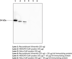
Vimentin is a cytoskeleton intermediate filament protein present in cells of mesenchymal origin, including leukocytes, endothelial cells, and smooth muscle cells.{42183} Each vimentin monomer contains a central α-helix that facilitates formation of the coil-coil dimer required for vimentin filament assembly.{42184} Vimentin is attached to nuclei, endoplasmic reticulum, and mitochondria, and has a role in positioning organelles in the cytosol. It regulates glial morphology, facilitates motility and directional migration of fibroblasts, and is critical to mechanotransduction of shear stress and maintenance of vascular endothelial integrity.{42183} Vimentin controls transport of LDL-derived cholesterol from lysosomes to esterification sites.{42185} It is an aggresome component, forming a cage-like structure around aggregated, undegraded proteins at the microtubule organizing center.{42186} Vimentin is subject to citrullination under high calcium concentrations, which can occur during macrophage apoptosis, and citrullinated vimentin has been shown to have a role in the production of anti-citrullinated protein antibodies (ACPAs).{31883,31884} ACPAs against citrullinated proteins, such as vimentin, are considered to be highly specific markers for rheumatoid arthritis and other autoimmune diseases.{31883} Cayman’s Vimentin Monoclonal Antibody (Clone 12E4) can be used for ELISA, immunoprecipitation, and Western blot applications. The antibody recognizes vimentin at ~54 kDa from human samples.
Brand:CaymanSKU:20197 - 100 µgAvailable on backorder
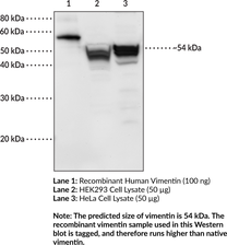
Vimentin is a cytoskeleton intermediate filament protein present in cells of mesenchymal origin, including leukocytes, endothelial cells, and smooth muscle cells.{42183} Each vimentin monomer contains a central α-helix that facilitates formation of the coil-coil dimer required for vimentin filament assembly.{42184} Vimentin is attached to nuclei, endoplasmic reticulum, and mitochondria, and has a role in positioning organelles in the cytosol. It regulates glial morphology, facilitates motility and directional migration of fibroblasts, and is critical to mechanotransduction of shear stress and maintenance of vascular endothelial integrity.{42183} Vimentin controls transport of LDL-derived cholesterol from lysosomes to esterification sites.{42185} It is an aggresome component, forming a cage-like structure around aggregated, undegraded proteins at the microtubule organizing center.{42186} Vimentin is subject to citrullination under high calcium concentrations, which can occur during macrophage apoptosis, and citrullinated vimentin has been shown to have a role in the production of anti-citrullinated protein antibodies (ACPAs).{31883,31884} ACPAs against citrullinated proteins, such as vimentin, are considered to be highly specific markers for rheumatoid arthritis and other autoimmune diseases.{31883} Cayman’s Vimentin Polyclonal Antibody can be used for Western blot and ELISA applications. The antibody recognizes vimentin at ~54 kDa from human samples.
Brand:CaymanSKU:25341 - 500 µgAvailable on backorder
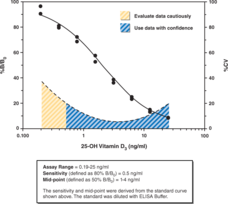
The primary synthesis of functional vitamin D begins in the skin, where a cholesterol by-product acted upon by UV light forms vitamin D3. Vitamin D3 is carried through the bloodstream, attached to the vitamin D binding protein, to the liver where it is converted to 25-hydroxy vitamin D3 (25(OH)D3). This product is then transported to the kidney where it is further converted to 1,25-dihydroxycholecalciferol (1,25(OH)2D3), the biologically active form. Active 1,25(OH)2D3 is very short-lived and is rapidly metabolized to its deactivated forms. Vitamin D Immunoassay: Accurate Vitamin D measurement in plasma or serum Cayman’s Vitamin D EIA Kit is a competitive assay that can be used for Vitamin D quantification in plasma or serum. Because of the short half-life of the biologically active 1,25(OH)D form, this Vitamin D assay primarily detects the more metabolically stable forms, 25(OH)D3 and 25(OH)D2. Accurate detection of these forms requires that they be displaced from the vitamin D binding protein prior to measurement. A simple, acetone-based purification protocol is included as the initial procedure of the Vitamin D assay kit for this purpose.
Brand:CaymanSKU:501050 - 480 solid wellsAvailable on backorder
The primary synthesis of functional vitamin D begins in the skin, where a cholesterol by-product acted upon by UV light forms vitamin D3. Vitamin D3 is carried through the bloodstream, attached to the vitamin D binding protein, to the liver where it is converted to 25-hydroxy vitamin D3 (25(OH)D3). This product is then transported to the kidney where it is further converted to 1,25-dihydroxycholecalciferol (1,25(OH)2D3), the biologically active form. Active 1,25(OH)2D3 is very short-lived and is rapidly metabolized to its deactivated forms. Vitamin D Immunoassay: Accurate Vitamin D measurement in plasma or serum Cayman’s Vitamin D EIA Kit is a competitive assay that can be used for Vitamin D quantification in plasma or serum. Because of the short half-life of the biologically active 1,25(OH)D form, this Vitamin D assay primarily detects the more metabolically stable forms, 25(OH)D3 and 25(OH)D2. Accurate detection of these forms requires that they be displaced from the vitamin D binding protein prior to measurement. A simple, acetone-based purification protocol is included as the initial procedure of the Vitamin D assay kit for this purpose.
Brand:CaymanSKU:501050 - 480 strip wellsAvailable on backorder
The primary synthesis of functional vitamin D begins in the skin, where a cholesterol by-product acted upon by UV light forms vitamin D3. Vitamin D3 is carried through the bloodstream, attached to the vitamin D binding protein, to the liver where it is converted to 25-hydroxy vitamin D3 (25(OH)D3). This product is then transported to the kidney where it is further converted to 1,25-dihydroxycholecalciferol (1,25(OH)2D3), the biologically active form. Active 1,25(OH)2D3 is very short-lived and is rapidly metabolized to its deactivated forms. Vitamin D Immunoassay: Accurate Vitamin D measurement in plasma or serum Cayman’s Vitamin D EIA Kit is a competitive assay that can be used for Vitamin D quantification in plasma or serum. Because of the short half-life of the biologically active 1,25(OH)D form, this Vitamin D assay primarily detects the more metabolically stable forms, 25(OH)D3 and 25(OH)D2. Accurate detection of these forms requires that they be displaced from the vitamin D binding protein prior to measurement. A simple, acetone-based purification protocol is included as the initial procedure of the Vitamin D assay kit for this purpose.
Brand:CaymanSKU:501050 - 96 solid wellsAvailable on backorder
The primary synthesis of functional vitamin D begins in the skin, where a cholesterol by-product acted upon by UV light forms vitamin D3. Vitamin D3 is carried through the bloodstream, attached to the vitamin D binding protein, to the liver where it is converted to 25-hydroxy vitamin D3 (25(OH)D3). This product is then transported to the kidney where it is further converted to 1,25-dihydroxycholecalciferol (1,25(OH)2D3), the biologically active form. Active 1,25(OH)2D3 is very short-lived and is rapidly metabolized to its deactivated forms. Vitamin D Immunoassay: Accurate Vitamin D measurement in plasma or serum Cayman’s Vitamin D EIA Kit is a competitive assay that can be used for Vitamin D quantification in plasma or serum. Because of the short half-life of the biologically active 1,25(OH)D form, this Vitamin D assay primarily detects the more metabolically stable forms, 25(OH)D3 and 25(OH)D2. Accurate detection of these forms requires that they be displaced from the vitamin D binding protein prior to measurement. A simple, acetone-based purification protocol is included as the initial procedure of the Vitamin D assay kit for this purpose.
Brand:CaymanSKU:501050 - 96 strip wellsAvailable on backorder
Brand:CaymanSKU:10007143 - 100 µLAvailable on backorder
Brand:CaymanSKU:10007143 - 500 µLAvailable on backorder
Brand:CaymanSKU:170120 - 100 µlAvailable on backorder
Brand:CaymanSKU:170120 - 500 µlAvailable on backorder
Brand:CaymanSKU:10007048 - 100 µlAvailable on backorder
Brand:CaymanSKU:10007048 - 500 µlAvailable on backorder