ELISA Kits
Showing 901–1050 of 3623 results
-
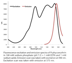
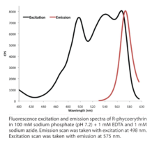
Goat anti-Rat IgG conjugated to SureLight® R-PE Antibody: Goat polyclonal anti-Rat IgG Dye: SureLight® R-phycoerythrin Excitation max. λ: 565>498 nm Emission max. λ: 578 nm Uses: Flow cytometry, cell-based assays, microarrays, and microplate applications
Brand:CaymanSKU:16652 - 100 µgAvailable on backorder
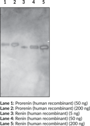
Cayman’s Goat Anti-Renin (human) Polyclonal Antibody is directed against the full-length renin (human recombinant) protein (Item No. 10006217). Renin is an aspartyl protease of approximately 40 kDa produced in the kidney by juxtaglomerular cells of the macula densa.{12331} Renin converts angiotensinogen into angiotensin I, activating a major arm of renal sodium and volume auto-regulation. Native human renin is a glycoprotein derived from preprorenin by sequential cleavage. The first cleavage releases the signal peptide (amino acids 1-23), forming the prorenin (44 kDa). The second cleavage removes the propeptide (amino acid 24-66), forming the functional renin protein (40 kDa). This antibody detects both the active renin and prorenin.
Brand:CaymanSKU:17623 - 1 eaAvailable on backorder
Goat F(Ab’)2 anti-Mouse IgG, H+L conjugated to SureLight® APC Antibody: Goat F(ab’)2 polyclonal anti-Mouse IgG Dye: SureLight® Allophycocyanin (APC) Excitation max. λ: 650 nm Emission max. λ: 660 nm Uses: Flow Cytometry and cell-based assays
Brand:CaymanSKU:16589 - 100 µgAvailable on backorder
Goat F(Ab’)2 anti-Mouse IgG, H+L conjugated to SureLight® R-PE Antibody: Goat F(Ab’)2 polyclonal anti-Mouse IgG Dye: SureLight® R-phycoerythrin Excitation max. λ: 565>498 nm Emission max. λ: 578 nm Uses: Flow cytometry, cell-based assays, microarrays, and microplate applications
Brand:CaymanSKU:16647 - 100 µgAvailable on backorder
Goat F(Ab’)2 anti-Mouse IgG, Fc-specific conjugated to SureLight® APC Antibody: Goat F(Ab’)2 polyclonal anti-Mouse IgG, Fc-specifc Dye: SureLight® Allophycocyanin (APC) Excitation max. λ: 650 nm Emission max. λ: 660 nm Uses: Flow Cytometry and cell-based assays
Brand:CaymanSKU:16590 - 100 µgAvailable on backorder
Goat F(Ab’)2 anti-Mouse IgG, Fc-specific conjugated to SureLight® R-PE Antibody: Goat F(Ab’)2 polyclonal anti-Mouse IgG, Fc-specific Dye: SureLight® R-phycoerythrin Excitation max. λ: 565>498 nm Emission max. λ: 578 nm Uses: Flow cytometry, cell-based assays, microarrays, and microplate applications
Brand:CaymanSKU:16648 - 100 µgAvailable on backorder
Goat F(Ab’)2 anti-Rat IgG conjugated to SureLight® APC Antibody: Goat F(Ab’)2 polyclonal anti-Rat IgG Dye: SureLight® Allophycocyanin (APC) Excitation max. λ: 650 nm Emission max. λ: 660 nm Uses: Flow Cytometry and cell-based assays
Brand:CaymanSKU:16594 - 100 µgAvailable on backorder
Goat F(Ab’)2 anti-Rat IgG conjugated to SureLight® R-PE Antibody: Goat F(Ab’)2 polyclonal anti-Rat IgG Dye: SureLight® R-phycoerythrin Excitation max. λ: 565>498 nm Emission max. λ: 578 nm Uses: Flow cytometry, cell-based assays, microarrays, and microplate applications
Brand:CaymanSKU:16653 - 100 µgAvailable on backorder
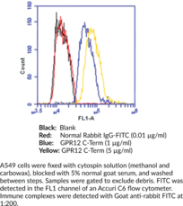
G protein-coupled receptor 12 (GPR12) is a high affinity receptor for sphingosine-1-phosphate, sphingosyl-phosphorylcholine and tyrosol that is expressed in brain, pituitary, ovary, and testis tissues.{22366,22367,22368,22364,22365} GPR12 plays a role in neuronal differentiation, neuronal growth and the formation of synaptic contacts.{22365,22367} Cayman’s GPR12 receptor (C-Term) polyclonal antibody can be used for flow cytometry and immunocytochemistry of GPR12 on human samples.
Brand:CaymanSKU:14266 - 1 eaAvailable on backorder
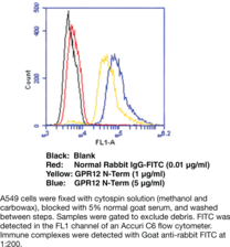
G protein-coupled receptor 12 (GPR12) is a high affinity receptor for sphingosine-1-phosphate, sphingosyl-phosphorylcholine and tyrosol that is expressed in brain, pituitary, ovary, and testis tissues.{22366,22367,22368,22364,22365} GPR12 plays a role in neuronal differentiation, neuronal growth and the formation of synaptic contacts.{22367,22365} Cayman’s GPR12 receptor (N-Term) polyclonal antibody can be used for flow cytometry and immunocytochemistry of GPR12 on human samples.
Brand:CaymanSKU:14267 - 1 eaAvailable on backorder
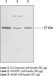
GPR17 is a G protein-coupled receptor that has been identified as a dualistic receptor recognizing signals from two unrelated chemical families: nucleotides and CysLTs.{16467} The deorphanization of GPR17 supports the suggested crosstalk between nucleotides and CysLTs during inflammation and injury. mRNA transcripts encoding this transmembrane receptor are most strongly expressed in the brain, kidney, and heart. Upon ischemic injury, GPR17 is upregulated in these tissues and its inhibition has been shown to decrease ischemic damage. This finding suggests GPR17 as a pharmacological target of ischemic injury.{16467} Cayman’s peptide affinity-purified polyclonal antibody recognizes the C-terminal region of human GPR17. This protein exists in two isoforms, differing by 28 amino acids at the receptor N-terminus. The longer form of the protein consists of 367 amino acids with a calculated molecular weight of 41 kDa. The human and rat proteins share 89% amino acid identity.{16467} Post-translational modifications may explain the observed SDS-PAGE gel-migration to 57 kDa on immunoblot.
Brand:CaymanSKU:10136 - 1 eaAvailable on backorder
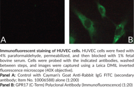
GPR17 is a G protein-coupled receptor that has been identified as a dualistic receptor recognizing signals from two unrelated chemical families: nucleotides and cysteinyl leukotrienes (CysLTs).{16467} The deorphanization of GPR17 supports the suggested crosstalk between nucleotides and CysLTs during inflammation and injury. mRNA transcripts encoding this transmembrane receptor are most strongly expressed in the brain, kidney, and heart. Upon ischemic injury, GPR17 is upregulated in these tissues and its inhibition has been shown to decrease ischemic damage. This finding suggests GPR17 as a pharmacological target of ischemic injury.{16467} Cayman’s GPR17 (C-Term) Polyclonal Antibody (Immunofluorescence) is a peptide affinity-purified polyclonal antibody that recognizes the C-terminal region of human GPR17. This protein exists in two isoforms, differing by 28 amino acids at the receptor N-terminus. The longer form of the protein consists of 367 amino acids with a calculated molecular weight of 41 kDa. The human and rat proteins share 89% amino acid identity.{16467}
Brand:CaymanSKU:17087 - 1 eaAvailable on backorder
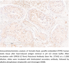
GPR18 is a G protein-coupled receptor (GPCR) that is expressed in lymphoid tissues, lung, brain, testis, and ovaries.{48187} It is coupled to the Gαi/o and Gαq/11 transduction pathways and is activated by the polyunsaturated fatty acid metabolite resolvin D2 (Item No. 10007279) and the endocannabinoid arachidonoyl glycine (NAGly; Item No. 90051). Resolvin D2-induced activation of GPR18 reduces inflammatory pain induced by formalin, carrageenan, and complete Freund’s adjuvant (CFA) in rats and reduces production of pro-inflammatory cytokines in mouse models of cecal ligation and puncture-induced sepsis, ocular infections, and ischemic injury.{42687,48188} NAGly-induced activation of GPR18 lowers intraocular pressure (IOP) in mice in a fatty acid amide hydrolase-dependent manner.{48189} GPR18 is also overexpressed and constitutively active in patient-derived metastatic melanoma tumor samples.{48190} Cayman’s GPR18 (C-Term) Polyclonal Antibody can be used for ELISA and immunohistochemistry (IHC) applications. The antibody recognizes the C-terminal region of GPR18 from human samples.
Brand:CaymanSKU:27342 - 500 µlAvailable on backorder
GPR31 is a 319 amino acid G protein-coupled receptor (GPCR) that acts as the receptor for the arachidonic acid metabolite 12-(S)-hydroxy-5,8,10,14-eicosatetraenoic acid (12(S)-HETE) (Item No. 34570). Binding of 12(S)-HETE to GPR31 leads to the activation of ERK1/2 (MAPK3/MAPK1), MEK, and NF-κB pathways. The dysregulation of GPR31 has been implicated in diseases such as arthritis, Alzheimer’s disease, and prostate cancer.{19754}
Brand:CaymanSKU:14083 - 1 eaAvailable on backorder
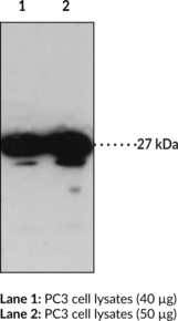
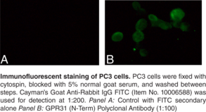
GPR31 is a 319 amino acid G protein-coupled receptor (GPCR) that acts as the receptor for the arachidonic acid (Item No. 90010) metabolite 12(S)-HETE (Item No. 34570). Binding of 12(S)-HETE to GPR31 leads to the activation of ERK1/2 (MAPK3/MAPK1), MEK, and NF-κB pathways. The dysregulation of GPR31 has been implicated in diseases such as arthritis, Alzheimer’s disease, and prostate cancer.{19754}
Brand:CaymanSKU:14033 - 1 eaAvailable on backorder
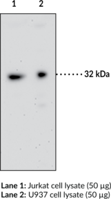
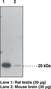
Many G protein-coupled receptor 35 (GPR35) ligands have recently been identified including kynurenic acid (Item No. 16762) and zaprinast (Item No. 110010421).{14771,14754} Multiple isoforms of this receptor have been reported and GPR35 mRNAs were detected in several tissues including intestine, lymphocytes, skeletal muscle, pancreatic β cells, and tumor cell lines.{14771,13309} Cayman’s GPR35 Polyclonal Antibody has been used successfully to detect this receptor in human and mouse intestine samples as well as in human, mouse, and porcine lymphocytes at 30 kDa on immunoblot.
Brand:CaymanSKU:10007660 - 1 eaAvailable on backorder
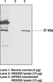
GPR55 is an orphan G protein-coupled receptor.{53310} GPR55 is expressed in the brain, large dorsal root ganglion neurons, and many peripheral tissues.{53311,17584} Endogenous agonists for GPR55 include lysophosphatidylinositol (EC50 = 1.2 µM in a β-arrestin-GFP biosensor assay) and the endocannabinoids anandamide (arachidonoyl ethanolamide; Item No. 90050) and 2-arachidonoyl glycerol (Item No. 62160; EC50s = 18.4 and 3.5 nM in GTPγS binding assays).{53310,53312} It is also activated by the cannabinoid Δ9-tetrahydrocannabinol (Δ9-THC; EC50 = 8 nM in a GTPγS binding assay).{53310,17581} GPR55 is expressed in β cells and pharmacological activation increases glucose-induced insulin release in wild-type mice and, to a lesser extent, in Gpr55 knockout mice.{53313} GPR55 expression is increased in the visceral adipose tissue of obese patients and, to a larger extent, in obese patients with type-2 diabetes.{53314} Activation of GPR55 increases the growth and invasiveness of cancer cells in vitro, and its expression in patient-derived tumors is positively correlated with a worse prognosis.{53313} GPR55 activation has also been associated with inhibition of osteoclast formation. Cayman’s GPR55 Polyclonal Antibody can be used for flow cytometry, immunofluorescence, and Western blot applications. The antibody recognizes GPR55 at 37 kDa from human and bovine samples. Post-translational modifications such as glycosylation may retard receptor electrophoretic migration such that the protein signal may be detected above 37 kDa.
Brand:CaymanSKU:10224 - 1 eaAvailable on backorder
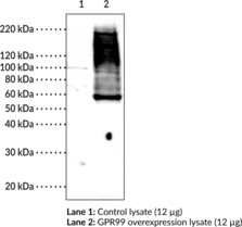
GPR99 is a G protein-coupled receptor (GPCR) that was originally identified as a purinergic receptor, P2Y15.{47265,47266} However, it is activated by leukotriene E4 (LTE4; Item No. 20410) in the low nanomolar range and by α-ketoglutarate in the high micromolar range.{23197,19519} It is found in the CNS, kidney, epididymis, and other tissues in the mouse, as well as in human kidney, nasal turbinates, and lung, among other tissues.{47267,47268} GPR99 knockout in mice prevents vascular leakage induced by LTE4 and epithelial cell mucin release in mouse nasal mucosa induced by LTE4 or A. alternata.{23197} In the kidney, under acid-base stress, it is involved with the regulation of carbonic acid and sodium chloride resorption through activation by α-ketoglutarate.{47267} GPR99 is also expressed in mouse retina, and axon growth is increased when it is activated by α-ketoglutarate in isolated mouse embryo retinal explants.{47269} Cayman’s GPR99 (C-Term) Polyclonal Antibody can be used for Western blot and immunohistochemistry (IHC) applications. The antibody recognizes the C-terminus of GPR99 from human samples.
Brand:CaymanSKU:27344 - 500 µLAvailable on backorder
Brand:CaymanSKU:703110 - 1 eaAvailable on backorder
GPX4 Assay Buffer (10x) has been tested and formulated to work exclusively with Cayman’s GPX4 Inhibitor Screening Assay Kit (Item No. 701880). Please visit GPX4 Inhibitor Screening Assay Kit (Item No. 701880) for the kit protocol, procedures, and product handling.
Brand:CaymanSKU:701881 - 1 eaAvailable on backorder
Cayman’s GPX4 Inhibitor Screening Assay Kit provides a robust and easy-to-use platform for identifying novel inhibitors of human GPX4, a negative regulator of the ferroptosis pathway. The assay measures GPX4 indirectly by a coupled reaction with glutathione reductase (GR). Oxidized glutathione (GSSG), produced upon reduction of hydroperoxide by GPX4, is recycled to its reduced state by GR and NADPH. The oxidation of NADPH to NADP+ is accompanied by a decrease in absorbance at 340 nm. The rate of decrease in A340 is directly proportional to GPX4 activity. The GPX4 inhibitor ML-162 is included as a positive control.
Brand:CaymanSKU:701880 - 96 wellsAvailable on backorder
Phospholipid hydroperoxide glutathione peroxidase (GPX4 or PhGPx) is one of four characterized selenium-dependent glutathione peroxidases.{7195} GPX4 is found primarily in the testis where it functions both to protect membrane phospholipids from oxidation in spermatids and as an insoluble structural protein in mature spermatozoa.{7195,7197} Three different post-translational forms of GPX4 have been characterized: mitochondrial, non-mitochondrial, and sperm nuclei specific selenoprotein (SnGPx).{12359} These monomeric GPX4 types migrate to 23, 20, and 34 kDa respectively.{7197,12359,12361,12360} GPX4 also forms a 69 kDa homotetramer.{12360} Cayman Chemical’s GPX4 polyclonal antibody can be used for western blot analysis of GPX4 on samples from mouse, rat, and pig, detecting both 20 kDa and 69 kDa bands. Sequence homology of the peptide antigen with that from other species indicates that the antibody is also expected to work on samples from human, bovine, canine, and chicken. Other applications for use of this antibody have not yet been tested.
Brand:CaymanSKU:10005258 - 1 eaAvailable on backorder
Brand:CaymanSKU:703210 - 1 eaAvailable on backorder
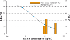
GH is a polypeptide hormone with a molecular weight of 23 kDa released from somatotropes of the anterior pituitary. It is regulated by several neurotransmitters and neuropeptides. Among other functions it plays an essential role in regulating body growth. Plasma GH levels in humans are markedly elevated in the perinatal period, in the range of 50 ng/ml, but decline to near-adult levels of 0.5-1.0 ng/ml within a few weeks. GH levels are slightly higher in women than in men, and are increased in both sexes in response to exercise. [Bertin Catalog No. A05104]
Brand:CaymanSKU:589601 - 96 wellsAvailable on backorder
Brand:CaymanSKU:32755 - 25 µLAvailable on backorder
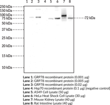
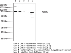
Glucose-regulated protein 78 kDa (GRP78), also known as heat shock 70 kDa protein 5 (HspA5) and immunoglobulin heavy chain-binding protein (BiP), is a glucose-regulated protein that is constitutively expressed in the lumen of the endoplasmic reticulum (ER).{36130,36131,36132} It is composed of two functional domains, an N-terminal nucleotide-binding domain that contains an ATP catalytic site and a C-terminal substrate binding domain that binds hydrophobic polypeptides.{36134} GRP78 functions as a molecular chaperone, assisting in the translocation of polypeptides from the cytosol into the ER, folding of nascent polypeptides, as well as refolding and preventing aggregation of misfolded proteins. It also plays a role in the ER-assisted degradation (ERAD) and unfolded protein response (UPR) pathways.{36137,36136} GRP78 chaperone activity is driven by an ATPase cycle that is regulated by ER-localized DnaJ-like protein co-factors and nuclear exchange factors.{36133,36135} Expression of GRP78 is upregulated in response to ER stress caused by viral and bacterial infections as well as various cancers.{36139} ER stress can also promote extracellular secretion of GRP78 leading to its anti-inflammatory functions in immunity.{36138} Cayman’s GRP78 Monoclonal Antibody can be used for Western blot and ELISA applications. The antibody recognizes GRP78 at ~72 kDa from human, mouse, and rat samples.
Brand:CaymanSKU:25690 - 100 µgAvailable on backorder
Glucose-regulated protein 78 kDa (GRP78), also known as heat shock 70 kDa protein 5 (HspA5) and immunoglobulin heavy chain-binding protein (BiP), is a glucose-regulated protein that is constitutively expressed in the lumen of the endoplasmic reticulum (ER).{36130,36131,36132} It is composed of two functional domains, an N-terminal nucleotide-binding domain that contains an ATP catalytic site and a C-terminal substrate binding domain that binds hydrophobic polypeptides.{36134} GRP78 functions as a molecular chaperone, assisting in the translocation of polypeptides from the cytosol into the ER, folding of nascent polypeptides, as well as refolding and preventing aggregation of misfolded proteins. It also plays a role in the ER-assisted degradation (ERAD) and unfolded protein response (UPR) pathways.{36137,36136} GRP78 chaperone activity is driven by an ATPase cycle that is regulated by ER-localized DnaJ-like protein co-factors and nuclear exchange factors.{36133,36135} Expression of GRP78 is upregulated in response to ER stress caused by viral and bacterial infections as well as various cancers.{36139} ER stress can also promote extracellular secretion of GRP78 leading to its anti-inflammatory functions in immunity.{36138} Cayman’s GRP78 Polyclonal Antibody can be used for Western blot and ELISA applications. The antibody recognizes GRP78 at ~72 kDa from human samples.
Brand:CaymanSKU:24533 - 500 µgAvailable on backorder
Glucose regulated protein 94 (GRP94) is a constitutively expressed endoplasmic reticulum (ER) lumenal protein that is up-regulated in response to cellular stress such as heat shock, oxidative stress or glucose depletion. GRP94 is thought to play a role in protein translocation to the ER, in their subsequent folding and assembly, and in regulating protein secretion.{15669} GRP94 also plays a role in antigen presentation by accessing the endogenous pathway and eliciting specific cytotoxic T lymphocyte (CTL) responses to chaperone bound peptides via the major histocompatibility complex (MHC) class I pathway.{15670} GRP94 is a member of the Hsp90 family of stress proteins and shares sequence homology with its cytosolic equivalent, Hsp90.{15671} Both Hsp90 and GRP94 are calcium binding proteins.{15672} Despite sharing 50% sequence homology over its N domains and complete conservation in its ligand binding domains with Hsp90, GRP94, and Hsp90 differ in their interactions with regulatory ligands as GRP94 has weak ATP binding and hydrolysis activity.{15673} GRP94 exists as a homodimer and the two subunits interact at two distinct intermolecular sites, C-terminal dimerization domains and the N-terminal interacts with the middle domain of opposing subunits.{15674} GRP94 contains a carboxy terminal KDEL (Lys-Asp-Glu-Leu) sequence which is believed to aid in its retention in the ER.{15675}
Brand:CaymanSKU:10011424 - 200 µgAvailable on backorder
Glucose regulated protein 94 (GRP94) is a constitutively expressed endoplasmic reticulum (ER) lumenal protein that is up-regulated in response to cellular stress such as heat shock, oxidative stress or glucose depletion. GRP94 is thought to play a role in protein translocation to the ER, in their subsequent folding and assembly, and in regulating protein secretion.{15669} GRP94 also plays a role in antigen presentation by accessing the endogenous pathway and eliciting specific cytotoxic T lymphocyte (CTL) responses to chaperone bound peptides via the major histocompatibility complex (MHC) class I pathway.{15670} GRP94 is a member of the Hsp90 family of stress proteins and shares sequence homology with its cytosolic equivalent, Hsp90.{15671} Both Hsp90 and GRP94 are calcium binding proteins.{15672} Despite sharing 50% sequence homology over its N domains and complete conservation in its ligand binding domains with Hsp90, GRP94, and Hsp90 differ in their interactions with regulatory ligands as GRP94 has weak ATP binding and hydrolysis activity.{15673} GRP94 exists as a homodimer and the two subunits interact at two distinct intermolecular sites, C-terminal dimerization domains and the N-terminal interacts with the middle domain of opposing subunits.{15674} GRP94 contains a carboxy terminal KDEL (Lys-Asp-Glu-Leu) sequence which is believed to aid in its retention in the ER.{15675}
Brand:CaymanSKU:10011424 - 50 µgAvailable on backorder
Glycogen synthase kinase 3 (GSK3) is a serine/threonine kinase that is involved in the regulation of many signaling pathways. To date, two isoforms have been identified: GSK3α and GSK3β. Specifically, GSK3β has been shown to play a key inhibitory role in both the insulin and Wnt signaling pathways. It has been suggested that Ser9 phosphorylation underlies the inhibition of GSK3β by insulin.
Brand:CaymanSKU:10009374 - 1 eaAvailable on backorder
Brand:CaymanSKU:703310 - 1 eaAvailable on backorder
Brand:CaymanSKU:32753 - 200 µgAvailable on backorder
Soluble guanylate cyclase is a heterodimeric enzyme, composed of α and β subunits, that synthesizes cGMP from GTP. The enzyme is activated by the binding of nitric oxide or carbon monoxide to the heme group of the enzyme.{1707} Chronic hypoxia upregulates soluble guanylate expression in rat lung.{7644} The α1 subunit contains 690-717 amino acids and has a molecular mass of 77-82 kDa.{4654,3536,3434} The cloned β1 subunit of guanylate cyclase from human, bovine, and rat sources contains 619 amino acids and has a molecular mass of approximately 70,000.{4654,3535,3531}
Brand:CaymanSKU:160895 - 1 eaAvailable on backorder
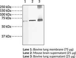
Soluble guanylate cyclase (sGC) is a heterodimeric hemoprotein and nitric oxide (NO) sensor composed of two subunits, α1 and β1.{59045,59046} The approximately 70 kDa sGC β1 subunit is encoded by GUCY1B3 in humans, ubiquitously expressed, and localized to the cytosol.{24381} The sGC histidine residue at position 105 is ligated to a ferrous heme that selectively binds NO to activate the C-terminal guanylate cyclase activity of the sGC heterodimer, catalyzing the synthesis of cGMP.{59045,59047} Knockdown of Gucy1B3 or expression of a heme-deficient sGC β1 subunit inhibits NO-induced reductions in blood pressure and platelet activation in mice, indicating a heme-dependent role for the sGC β1 subunit in blood pressure regulation.{59048} Cayman’s Guanylate Cyclase β1 subunit (soluble) Polyclonal Antibody can be used for immunohistochemistry (IHC) and Western blot (WB) applications. The antibody recognizes the sGC β1 subunit from human, bovine, and rat samples.
Brand:CaymanSKU:160897 - 1 eaAvailable on backorder
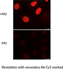
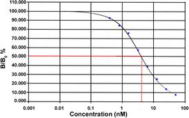
DNA double stand break (DSB) are leading to the early phosphorylation of histone H2AX. Phosphorylated γ-H2AX is the first step in the recruitment of the DNA repair protein at the foci site. They are consequently very good biomarkers of DSB as well as check point for cell cycle arrest following DSB. Measuring γ-H2AX phosphorylation has become a gold standard in DNA damage and repair for DSB events.
Brand:CaymanSKU:25781 - 100 µlAvailable on backorder
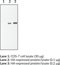
The HA epitope is an expression tag commonly used in protein expression experiments. Compared to its corresponding native protein, a HA-tagged protein will migrate at a larger size on SDS-PAGE, which is equivalent to the size of the epitope tag. The size of the HA epitope may vary depending on the expression vector being utilized, but usually ranges from 3 to 5 kDa. Cayman Chemical’s HA polyclonal antibody will recognize proteins expressed with both N-terminal and C-terminal HA tags.
Brand:CaymanSKU:162200 - 1 eaAvailable on backorder
Brand:CaymanSKU:10009330 - 1 eaAvailable on backorder
Cayman’s Histone Acetyltransferase (HAT) Inhibitor Screening Assay Kit provides a fast, fluorescence-based method for evaluating PCAF HAT inhibitors. The procedure requires only three easy steps, all performed in the same microwell plate. Cayman’s Histone Acetyltransferase (HAT) Inhibitor Screening Assay Kit provides a fast, fluorescence-based method for evaluating PCAF HAT inhibitors. The procedure requires only three easy steps, all performed in the same microwell plate. In the first step of the protocol, HAT is incubated with acetyl-CoA and the histone H3 peptide. During this time, HAT catalyzes the enzymatic transfer of acetyl groups from acetyl-CoA to the H3 peptide producing an acetylated peptide and CoASH. Following addition of isopropanol to stop the enzymatic reaction, CPM is added to the wells of the plate. CPM reacts with the free thiol groups present on CoASH forming a highly fluorescent product that is detected using excitation and emission wavelengths of 360-390 and 450-470 nm, respectively.
Brand:CaymanSKU:10006515 - 96 wellsAvailable on backorder
Brand:CaymanSKU:700012 - 1 mlAvailable on backorder
Hyperpolarization-activated cation channels of the HCN gene family, such as HCN1, play a crucial role in the regulation of cell excitability. Importantly, they contribute to spontaneous rhythmic activity in both the heart and brain.{17645}
Brand:CaymanSKU:13705 - 100 µgAvailable on backorder
Hyperpolarization-activated cation channels of the HCN gene family contribute to spontaneous rhythmic activity in both the heart and the brain.{17645}
Brand:CaymanSKU:13707 - 100 µgAvailable on backorder
Hyperpolarization-activated cation channels of the HCN gene family contribute to spontaneous rhythmic activity in both the heart and the brain.{17645}
Brand:CaymanSKU:13708 - 100 µgAvailable on backorder
Ion channels are integral membrane proteins that help establish and control the small voltage gradient across the plasma membrane of living cells by allowing the flow of ions down their electrochemical gradient.{17533} They are present in the membranes that surround all biological cells and their main function is to regulate the flow of ions across this membrane. Whereas some ion channels permit the passage of ions based on charge, others conduct based on a ionic species, such as sodium or potassium. Furthermore, in some ion channels, the passage is governed by a gate which is controlled by chemical or electrical signals, temperature, or mechanical forces. There are a few main classifications of gated ion channels. There are voltage-gated ion channels, ligand-gated, other gating systems, and finally those that are classified differently, having more exotic characteristics. The first are voltage-gated ion channels which open and close in response to membrane potential. These are then seperated into sodium, calcium, potassium, proton, transient receptor, and cyclic nucleotide-gated channels, each of which is responsible for a unique role. Ligand-gated ion channels are also known as ionotropic receptors and they open in response to specific ligand molecules binding to the extracellular domain of the receptor protein. The other gated classifications include activation and inactivation by second messengers, inward-rectifier potassium channels, calcium-activated potassium channels, two-pore-domain potassium channels, light-gated channels, mechano-sensitive ion channels, and cyclic nucleotide-gated channels. Finally, the other classifications are based on less normal characteristics such as two-pore channels and transient receptor potential channels.{17535} Hyperpolarization-activated cation channels of the HCN gene family contribute to spontaneous rhythmic activity in both the heart and the brain.{17645}
Brand:CaymanSKU:13709 - 100 µgAvailable on backorder
Brand:CaymanSKU:10006389 - 1 eaAvailable on backorder
Nucleosomes, which fold chromosomal DNA, contain two molecules each of the core histones H2A, H2B, H3, and H4. Almost two turns of DNA are wrapped around this octameric core, which represses transcription.{12364} The histone amino termini extend from the core, where they can be modified post-translationally by acetylation, phosphorylation, ubiquitination, and methylation, affecting their charge and function. Acetylation of the ε-amino groups of specific histone lysines is catalyzed by histone acetyltransferases (HATs) and correlates with an open chromatin structure and gene activation. Histone deacetylases (HDACs) catalyze the hydrolytic removal of acetyl groups from histone lysine residues and correlates with chromatin condensation and transcriptional repression.{12365,12366} Therefore, HDAC inhibition results in transcriptional activation through the conformational relaxation of DNA. Changes in the transcription of key genes has linked HDAC inhibitors to blocking angiogenesis and cell cycling, and promoting apoptosis and differentiation. By targeting these key components of tumor proliferation, HDAC inhibitors are currently being explored as potential anticancer agents.{12367,12368,10618} Cayman’s HDAC Cell-Based Assay Kit provides an easy tool for studying HDAC activity modulators in whole cells. By using a cell-permeable HDAC substrate, the activity of various protein lysine-specific deacetylases including HDAC1-containing complexes can be measured in intact cells in a simple and homogenous manner. The fluorescence of the deacetylated reaction product can be analyzed using a plate reader or a fluorometer with excitation wavelengths of 340-360 nm and emission wavelengths of 440-465 nm. An HDAC inhibitor, trichostatin A, is included for checking specificity of the HDAC reaction. This assay parallels Cayman’s HDAC Activity Assay Kit (Item No. 10011563), which uses a nuclear extract rather than whole cells for the assay. Together, both assays will help to identify whether an inhibitor/activator has a direct effect on the enzyme.
Brand:CaymanSKU:600150 - 96 wellsAvailable on backorder
Histone deacetylases (HDACs) catalyze the hydrolytic removal of acetyl groups from histone lysine residues resulting in chromatin condensation and transcriptional repression of chromosomal DNA. Thus, HDAC inhibition allows the conformation of DNA to be relaxed and transcriptional activation to ensue. Transcriptional repression of key genes has linked HDAC activity to cell growth and tumor development. Cayman’s HDAC Activity Assay Kit provides a fast, fluorescence-based method for measuring Class I and II HDAC activity that eliminates radioactivity, extraction, or chromatography. The procedure requires only two easy steps, both performed in the same microplate. The fluorescent reaction product is analyzed using a plate reader with excitation wavelengths of 340-360 nm and emission wavelengths of 440-465 nm.
Brand:CaymanSKU:10011563 - 96 wellsAvailable on backorder
Brand:CaymanSKU:10011618 - 1 eaAvailable on backorder
Histone deacetylases (HDACs) catalyze the hydrolytic removal of acetyl groups from histone lysine residues resulting in chromatin condensation and transcriptional repression of chromosomal DNA. Thus, HDAC inhibition allows the conformation of DNA to be relaxed and transcriptional activation to ensue. Changes in the transcription of key genes have linked HDAC inhibitors to blocking angiogenesis and cell cycling, and promoting apoptosis and differentiation. Cayman’s HDAC1 Inhibitor Screening Assay Kit provides a fast, fluorescence-based method for screening HDAC1 inhibitors. The procedure requires only two easy steps, both performed in the same microplate. The fluorescent reaction product is easily analyzed using a fluorometer with excitation wavelengths of 340-360 nm and emission wavelengths of 440-465 nm. Sufficient purified HDAC1 is provided for 100 tests.
Brand:CaymanSKU:10011564 - 96 wellsAvailable on backorder
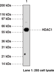
Histone deacetylase (HDAC) and histone acetyltransferase (HAT) are enzymes that regulate transcription by selectively deacetylating or acetylating the ε-amino groups of lysines located near the amino termini of core histone proteins.{17241} Eleven members of HDAC family have been identified in the past several years.{17242,17240} These HDAC family members are divided into two classes, I and II. HDAC1 is a Class I HDAC which is related to the yeast HDAC Rpd3.{14468} It is primarily localized to the nucleus with ubiquitous distribution throughout human cell lines and tissues. By modifying chromatin structure and other non-histone proteins, HDACs play important roles in controlling complex biological events, including cell development, differentiation, programmed cell death, angiogenesis, and inflammation. Considering these major roles, it is conceivable that dysregulation of HDACs and subsequent imbalance of acetylation and deacetylation may be involved in the pathogenesis of various diseases, including cancer and inflammatory diseases.{14468}
Brand:CaymanSKU:13491 - 1 eaAvailable on backorder
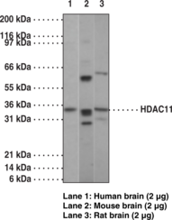
Histone deacetylase (HDAC) and histone acetyltransferase (HAT) are enzymes that regulate transcription by selectively deacetylating or acetylating the ε-amino groups of lysines located near the amino termini of core histone proteins.{17343} Eleven members of HDAC family have been identified in the past several years.{17241,17242} These HDAC family members are divided into two classes, I and II.The newest memeber of this family, HDAC11, has been cloned by Gao et al.{17240} HDAC11 transcripts were limited to kidney, heart, brain, skeletal muscle, and testis. These results suggested that each member of the HDAC family exhibits a different, individual substrate specificity and function in vivo.
Brand:CaymanSKU:13504 - 1 eaAvailable on backorder
Histone deacetylase (HDAC) and histone acetyltransferase (HAT) are enzymes that regulate transcription by selectively deacetylating or acetylating the ε-amino groups of lysines located near the amino termini of core histone proteins.{17241} Eleven members of HDAC family have been identified in the past several years.{17242,17240} These HDAC family members are divided into two classes, I and II. HDAC3 is a Class I HDAC which is related to the yeast HDAC Rpd3.{14468} It is primarily localized to the nucleus with ubiquitous distribution throughout human cell lines and tissues. By modifying chromatin structure and other non-histone proteins, HDACs play important roles in controlling complex biological events, including cell development, differentiation, programmed cell death, angiogenesis, and inflammation. Considering these major roles, it is conceivable that dysregulation of HDACs and subsequent imbalance of acetylation and deacetylation may be involved in the pathogenesis of various diseases, including cancer and inflammatory diseases.{14468}
Brand:CaymanSKU:13493 - 1 eaAvailable on backorder
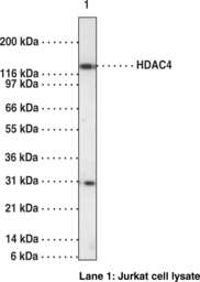
Histone deacetylase (HDAC) and histone acetyltransferase (HAT) are enzymes that regulate transcription by selectively deacetylating or acetylating the ε-amino groups of lysines located near the amino termini of core histone proteins.{17241} Eleven members of HDAC family have been identified in the past several years.{17242,17243} These HDAC family members are divided into two classes, I and II. HDAC4 is a member of the class II family which also includes HDACs 5, 6, and 7, the molecular weights of which are all about two-fold larger than those of the class I members.
Brand:CaymanSKU:13494 - 1 eaAvailable on backorder
Histone deacetylase (HDAC) and histone acetyltransferase (HAT) are enzymes that regulate transcription by selectively deacetylating or acetylating the ε-amino groups of lysines located near the amino termini of core histone proteins.{17240} Eleven members of the HDAC family have been identified in the past several years.{17241,17242} These HDAC family members are divided into two classes, I and II. HDAC6 is a Class II HDAC that can shuttle between the nucleus and cytoplasm, suggesting potential extranuclear functions by regulating the acetylation status of non-histone substrates. By modifying chromatin structure and other non-histone proteins, HDACs play important roles in controlling complex biological events, including cell development, differentiation, programmed cell death, angiogenesis, and inflammation.{14467,14468} Considering these major roles, it is conceivable that dysregulation of HDACs and subsequent imbalance of acetylation and deacetylation may be involved in the pathogenesis of various diseases, including cancer and inflammatory diseases.{14467}
Brand:CaymanSKU:13499 - 1 eaAvailable on backorder
Histone deacetylase 7 (HDAC7) is a thymus-specific, intracellular transcription factor belonging to the class IIa HDAC family. Reports suggest that dephosphorylation and nuclear localization of HDAC7 is promoted by myosin phosphatase, thus regulating the repression of Nur77 and inhibition of apoptosis in CD4+/CD8+ double-positive thymocytes. HDAC7 plays a specific role in maintaining vascular integrity by repressing the expression of matrix metalloproteinase (MMP) 10. It promotes repression mediated by transcriptional corepressor NCOR2 and is an efficient corepressor of the androgen receptor (AR). It is also responsible for the deacetylation of lysine residues on the N-terminal part of the core histones (H2A, H2B, H3, and H4).
Brand:CaymanSKU:13500 - 1 eaAvailable on backorder
Brand:CaymanSKU:700232 - 1 eaAvailable on backorder
Brand:CaymanSKU:700231 - 1 eaAvailable on backorder
Human HDAC8 is a class I HDAC and has been identified in a variety of human cancer tissues. Cayman’s HDAC8 Inhibitor Screening Assay provides a convenient fluorescence-based method for screening HDAC8 inhibitors. The procedure requires only two easy steps, both performed in the same microplate. The fluorescent reaction product is analyzed with an excitation wavelength between 350-360 nm and an emission wavelength between 450-465 nm. Sufficient HDAC8 is provided for 100 tests.
Brand:CaymanSKU:700230 - 96 wellsAvailable on backorder
Brand:CaymanSKU:32978 - 100 µlAvailable on backorder
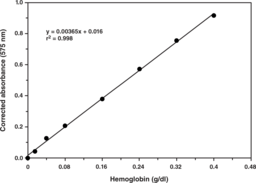
Hemoglobin (Hb or Hgb) is a globular protein found primarily in erythrocytes that carries oxygen from the lungs to tissue where it releases oxygen and then returns carbon dioxide (CO2) from the tissue to the lungs. Beyond binding and transporting oxygen, hemoglobin also binds CO2, carbon monoxide (CO), and nitric oxide (NO). Cayman’s Hemoglobin Colorimetric Assay provides a quick, reliable method for determining total hemoglobin concentration in a variety of biological samples, including blood, tissue homogenates, and cell lysates. Unlike the universally accepted reference method of hemoglobin determination, which uses potassium cyanide as a reagent and commonly under-estimates hemoglobin levels, Cayman’s Hemoglobin Assay utilizes an optimized detection reagent which is non-toxic, reporting accurate measurements of total hemoglobin concentrations with an absorbance between 560-590 nm.
Brand:CaymanSKU:700540 - 2 x 96 wellsAvailable on backorder
Hepsin is a type II membrane-associated protein that has an extracellular proteolytic domain and exhibits low sequence homology to other known proteases.{9602,9603} Hepsin overexpression is observed in prostate, breast, kidney, and ovarian cancers and due to low homology to other known proteases may provide a unique target for pharmacological therapy.{9067,9600,9601,9599,9604} Hepsin is necessary for cell growth in vitro and may play a role in metastatic expansion by factor VII activation.{9598,9606,9599} Cayman Chemical’s hepsin polyclonal antibody can be used for WB and IHC (formalin-fixed paraffin-embedded tissue) analysis for hepsin on samples of human origin. Other applications for use of this antibody have not yet been tested.
Brand:CaymanSKU:100022 - 500 µlAvailable on backorder
Human epidermal growth factor receptor 2 (HER2), also known as ErbB2 and Neu, is a cell surface receptor and member of the EGF family of receptor tyrosine kinases that is encoded by ERBB2 in humans.{56034} It is a transmembrane receptor composed of a C-terminal intracellular tyrosine kinase domain, a transmembrane lipophilic segment, and an N-terminal extracellular domain, that is expressed at low levels in various epithelial tissues, as well as breast, lung, kidney, ovary, placenta, and the gastrointestinal tract.{56035} Unlike other EGF receptors, HER2 does not bind ligands or undergo a conformational change in its extracellular domain for activation. HER2 is activated upon heterodimerization with HER3 or HER4, which stabilizes ligand binding to HER3 and HER4, or homodimerization and enhances kinase-mediated activation of the MAPK and PI3K cellular signaling pathways.{56034,56035} Truncated forms of HER2 with constitutive oncogenic activity can be generated by proteolytic cleavage of the extracellular domain.{56035} ERBB2 is overexpressed in 12 to 15% of breast cancer tumors and is associated with accelerated growth rate, increased rate of recurrence, and poor overall survival.{56035} Various mutations in ERBB2, with or without gene amplification, have been found in prostate, colon, bladder, breast, lung, and colorectal tumors, as well as metastatic cutaneous squamous small cell carcinomas.{56037,56036} Cayman’s HER2/ErbB2 (C-Term) Monoclonal Antibody can be used for immunohistochemistry (IHC) and Western blot (WB) applications.
Brand:CaymanSKU:32191 - 1 mlAvailable on backorder
Human epidermal growth factor receptor 2 (HER2), also known as ErbB2 and Neu, is a cell surface receptor and member of the EGF family of receptor tyrosine kinases that is encoded by ERBB2 in humans.{56034} It is a transmembrane receptor composed of a C-terminal intracellular tyrosine kinase domain, a transmembrane lipophilic segment, and an N-terminal extracellular domain, that is expressed at low levels in various epithelial tissues, as well as breast, lung, kidney, ovary, placenta, and the gastrointestinal tract.{56035} Unlike other EGF receptors, HER2 does not bind ligands or undergo a conformational change in its extracellular domain for activation. HER2 is activated upon heterodimerization with HER3 or HER4, which stabilizes ligand binding to HER3 and HER4, or homodimerization and enhances kinase-mediated activation of the MAPK and PI3K cellular signaling pathways.{56034,56035} Truncated forms of HER2 with constitutive oncogenic activity can be generated by proteolytic cleavage of the extracellular domain.{56035} ERBB2 is overexpressed in 12 to 15% of breast cancer tumors and is associated with accelerated growth rate, increased rate of recurrence, and poor overall survival.{56035} Various mutations in ERBB2, with or without gene amplification, have been found in prostate, colon, bladder, breast, lung, and colorectal tumors, as well as metastatic cutaneous squamous small cell carcinomas.{56037,56036} Cayman’s HER2/ErbB2 (C-Term) Monoclonal Antibody can be used for immunohistochemistry (IHC) and Western blot (WB) applications.
Brand:CaymanSKU:32191 - 100 µlAvailable on backorder
Brand:CaymanSKU:32902 - 100 µlAvailable on backorder
Brand:CaymanSKU:32917 - 100 µlAvailable on backorder
Brand:CaymanSKU:32918 - 100 µlAvailable on backorder
Hedgehog acyltransferase-like (HHATL) is the mammalian homolog of yeast Gup1 that is encoded by the HHATL gene in humans.{53526} HHATL is a membrane-bound O-acyltransferase (MBOAT) superfamily member and contains multiple transmembrane domains common to this family but lacks acyltransferase function due to a single amino acid substitution of histidine for leucine at position 447.{53526,27302} It is localized to the endoplasmic reticulum (ER) and is expressed in the heart, skeletal muscle, and brain.{53526,27300,53527} HHATL colocalizes with sonic hedgehog (Shh) and is a negative regulator of Shh N-terminal palmitoylation, a post-translational modification that is critical for Shh signaling in neural development and embryogenesis.{27300} Hhatl-/- neonatal mice fail to develop proper suckling ability, leading to malnutrition and death by postnatal day 14.{53528} HHATL expression is decreased in six nasopharyngeal carcinoma cell lines, as well as in tissue isolated from patients with nasopharyngeal carcinoma or skin squamous cell carcinoma.{53529,53530} Cayman’s HHATL Polyclonal Antibody can be used for flow cytometry (FC), immunofluorescence (IF), and Western blot applications. The antibody recognizes HHATL at ~60 kDa from human samples.
Brand:CaymanSKU:15648 - 1 eaAvailable on backorder
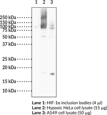
Hypoxia-inducible factor-1α (HIF-1α) is a transcription factor subunit that belongs to the basic helix-loop-helix PER-ARNT-SIM (bHLH-PAS) protein family.{11044,46550} It contains bHLH and PAS domains that mediate DNA binding and heterodimerization with the HIF-1β subunit, an oxygen-dependent degradation (ODD) domain that is hydroxylated by prolyl hydroxylase in the presence of oxygen to target HIF-1α for proteasomal degradation, and N- and C-terminal transactivation domains responsible for regulating the expression of HIF-1 target genes.{46550,46551} Under hypoxic conditions, HIF-1α is stabilized, accumulates in the cytoplasm, and is translocated to the nucleus where it forms a heterodimer with HIF-1β and induces the expression of genes involved in maintaining cellular oxygen homeostasis.{46550,10948,11044,12428} It is also involved in angiogenesis, glucose utilization, and pH regulation under hypoxic conditions, including in the tumor microenvironment.{17023} HIF-1α is overexpressed in a variety of cancer cell lines where it promotes survival of cancer cells and increases invasiveness under hypoxic conditions and, in vivo, overexpression is associated with aggressiveness and progression of various cancers and poor disease-free survival.{9833,46552,17023,46553} Homozygous knockout of HIF-1α is embryonic lethal due to disruptions in vascular development but conditional knockout models have demonstrated a role for HIF-1α in inflammation, immunity, and osteogenesis.{17023} Cayman’s HIF-1α Monoclonal Antibody can be used for Western blot and immunocytochemistry applications. The antibody recognizes HIF-1α at 93 kDa from human and mouse samples.
Brand:CaymanSKU:27227 - 100 µgAvailable on backorder
Hypoxia-inducible factor-1α (HIF-1α) is a transcription factor that accumulates under low-oxygen conditions.{10948,11044} Following hypoxic stimulus and cytoplasmic accumulation, HIF-1α migrates to the nucleus where with other transcription factors, it drives the production of stress-adaptive proteins. This response is essential for maintenance of normal oxidative physiology, however overexpression in cancer cells promotes tumor survival.{11044,9833,9834,9835,10695} HIF-1α is an endothelial PAS domain protein 1 (EPAS-1) family member with phosphorylation dependent activities.{11043,10898} Cayman Chemical’s HIF-1α polyclonal antibody can be used for WB analysis for HIF-1α on samples from human, mouse, and simian. It is not suitable for ICC or IP. Other applications for use of this antibody have not yet been tested.
Brand:CaymanSKU:10006421 - 1 eaAvailable on backorder
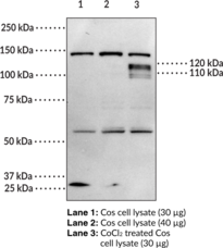
Hypoxia-inducible factor-1α (HIF-1α) is a transcription factor subunit that belongs to the basic helix-loop-helix PER-ARNT-SIM (bHLH-PAS) protein family.{11044,46550} It contains bHLH and PAS domains that mediate DNA binding and heterodimerization with the HIF-1β subunit, an oxygen-dependent degradation (ODD) domain that is hydroxylated by prolyl hydroxylase in the presence of oxygen to target HIF-1α for proteasomal degradation, and N- and C-terminal transactivation domains responsible for regulating the expression of HIF-1 target genes.{46550,46551} Under hypoxic conditions, HIF-1α is stabilized, accumulates in the cytoplasm, and is translocated to the nucleus where it forms a heterodimer with HIF-1β and induces the expression of genes involved in maintaining cellular oxygen homeostasis.{11044,46550,10948,12428} It is also involved in angiogenesis, glucose utilization, and pH regulation under hypoxic conditions, including in the tumor microenvironment.{17023} HIF-1α is overexpressed in a variety of cancer cell lines where it promotes survival of cancer cells and increases invasiveness under hypoxic conditions and, in vivo, overexpression is associated with aggressiveness and progression of various cancers and poor disease-free survival.{17023,9833,46552,46553} Homozygous knockout of HIF-1α is embryonic lethal due to disruptions in vascular development but conditional knockout models have demonstrated a role for HIF-1α in inflammation, immunity, and osteogenesis.{17023} Cayman’s HIF-1α (human) Monoclonal Antibody can be used for immunohistochemistry (IHC) and Western blot (WB) applications. The antibody recognizes HIF-1α at approximately 120 kDa from human samples.
Brand:CaymanSKU:32196 - 100 µlAvailable on backorder
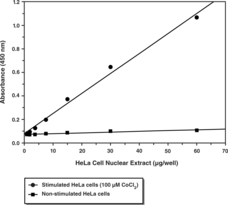
The HIF (hypoxia-inducible factor) transcription factor is a member of the basic-helix-loop-helix (bHLH) family of transcription factors and plays an important role in maintaining cellular oxygen homeostasis.{11044,12428} HIF-1α has emerged as an important drug target in breast and prostate cancer, cardiovascular disease, and ischemia.{10485,10724,11835} Cayman’s HIF-1α Transcription Factor Assay is a non-radioactive, sensitive method for detecting specific transcription factor DNA binding activity in nuclear extracts and whole cell lysate. A 96-well enzyme-linked immunosorbent assay (ELISA) replaces the cumbersome radioactive electrophoretic mobility shift assay (EMSA). A specific double stranded DNA (dsDNA) consensus sequence containing the HIF-1α response element is immobilized to the wells of a 96-well plate. HIF-1α contained in a nuclear extract or whole cell lysates, binds specifically to the HIF-1α response element. The HIF transcription factor complex is detected by addition of a specific primary antibody directed against HIF-1α. A secondary antibody conjugated to HRP is added to provide a sensitive colorimetric readout at 450 nm.
Brand:CaymanSKU:10006910 - 96 wellsAvailable on backorder
Recombinant proteins are routinely labeled with affinity tags, such as hexahistidine (6X-His), GST and FLAG, to facilitate both their detection and purification. 6X-His tags are often the tag of choice due to their small size, less potential to interfere in protein folding, weak immunogenicity, and high affinity for Ni2+. Cell lysates and samples at different stages of purification are generally analyzed by SDS-PAGE, a process which can add several days to the purification and analysis protocol. A semi-quantitative screening assay provides the ability to rapidly assess the levels His-tagged proteins at each stage of the expression and purification process. This permits the user to quickly monitor expression efforts, as well as follow protein loss or enrichment at each purification step. Cayman’s His-Tag Detection ELISA Kit is competitive assay designed for the rapid, semi-quantitative screening of cell lysates and affinity column fractions for His-tagged proteins. It is intended to serve as a substitute for SDS-PAGE, thereby expediting the screening of affinity column fractions.
Brand:CaymanSKU:10012445 - 96 solid wellsAvailable on backorder
Recombinant proteins are routinely labeled with affinity tags, such as hexahistidine (6X-His), GST and FLAG, to facilitate both their detection and purification. 6X-His tags are often the tag of choice due to their small size, less potential to interfere in protein folding, weak immunogenicity, and high affinity for Ni2+. Cell lysates and samples at different stages of purification are generally analyzed by SDS-PAGE, a process which can add several days to the purification and analysis protocol. A semi-quantitative screening assay provides the ability to rapidly assess the levels His-tagged proteins at each stage of the expression and purification process. This permits the user to quickly monitor expression efforts, as well as follow protein loss or enrichment at each purification step. Cayman’s His-Tag Detection ELISA Kit is competitive assay designed for the rapid, semi-quantitative screening of cell lysates and affinity column fractions for His-tagged proteins. It is intended to serve as a substitute for SDS-PAGE, thereby expediting the screening of affinity column fractions.
Brand:CaymanSKU:10012445 - 96 strip wellsAvailable on backorder
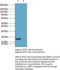
His-tag Monoclonal Antibody is a probe for the immunochemical detection of histidine tags on recombinant proteins. Recombinant proteins are commonly labeled with affinity tags, such as 6-10 histidine residues (6x-10x His), to facilitate both their detection and purification.{59520} Poly-His tags are commonly utilized because of their small size, low potential to interfere in protein folding or activity, weak immunogenicity, and high affinity for transition metal ion matrices, such as Ni2+, used in immobilized metal-affinity chromatography (IMAC) for protein purification.{59521,59520} Cayman’s His-tag Monoclonal Antibody is comprised of rabbit IgG variable domains and the human IgG1 constant domain. This antibody can be used for ELISA and Western blot (WB) applications. It recognizes proteins containing 6x or 10x His tags fused to either the N- or C-terminus and is recognized by anti-human IgG secondary antibodies.
Brand:CaymanSKU:32187 - 100 µgAvailable on backorder
Histamine (1H-imidazole 4-ethaneamine) is a component of the storage granules of myeloid cells including basophils{4343} and mast cells.{4178} Histamine secretion from gastric serosal cells contributes to the stimulation of gastric acid secretion. The clinical importance of histamine in the pathogenesis of allergy and asthma has long been appreciated.{2073} Together with the cysteinyl-leukotrienes, histamine contributes to the airway constriction, edema, vascular congestion, and inflammatory cell recruitment seen in acute allergic respiratory disease. Bacterial decarboxylation of the amino acid histidine in fresh fish produces histamine, which can be used as a health hazard biomarker indicative of bacterial contamination. The measurement of histamine levels is therefore of importance in fields as diverse as food quality inspection, pharmaceutical development, and clinical diagnostics. Our Histamine EIA kit is a derivitization-amplified competitive enzyme immunoassay which detects histamine within the range from 400 pg/ml to 50,000 pg/ml. The assay can be used for the analysis of histamine in blood (plasma or serum) without extraction or purification. The use of other sample types may require further processing or purification of the sample. [Bertin Catalog No. A05890]
Brand:CaymanSKU:589651 - 96 wellsAvailable on backorder
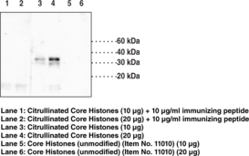
Citrullinated histone H1.4 is a citrullinated form of histone H1.4, a nuclear protein that interacts with linker DNA regions between nucleosomes to provide nucleosome core structure stabilization.{52742} Histone H1 subtypes are comprised of a short N-terminal domain, a central globular domain necessary for DNA interaction, and long C-terminal domain that determines subtype binding properties by its variability. Histone H1.4 is expressed in most somatic cell types and is essential for survival of T47D and MCF-7 breast cancer cells and MCF-10A breast epithelial cells.{52743,52744,52745} It is subject to citrullination by protein-arginine deiminase 4 (PAD4) at arginine 54, which corresponds to arginine 53 of the immunizing peptide for Cayman’s Histone H1.4 (Citrullinated R53) Polyclonal Antibody. Citrullination releases histone 1 from chromatin, leading to chromatin decondensation in mouse embryonic stem cells.{52746} Cayman’s Histone H1.4 (Citrullinated R53) Polyclonal Antibody can be used for Western blot applications. The antibody recognizes citrullinated histone H1, and not unmodified H1, at 35 kDa from human samples.
Brand:CaymanSKU:18073 - 1 eaAvailable on backorder
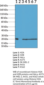
Histone H2A is a nuclear protein and a component of the nucleosome core, a basic unit of chromatin, that is essential for organizing genomic DNA in eukaryotic nuclei.{57056} It is a globular protein that contains an unstructured N-terminal tail that extends outside of the nucleosome core and is subject to a variety of post-translational modifications (PTMs), including ubiquitination, acetylation, methylation, and phosphorylation, which function as epigenetic regulators of transcription.{18136,57056} Histone H2A contains a C-terminal tail that, in contrast to other core histones, also extends outside of the nucleosome core and is highly variable, yielding a number of H2A variants that are subject to PTMs and differentially regulate nucleosome stability and chromatin structure.{58069} Serum histone H2A autoantibodies have been found in patients with drug-induced or spontaneous systemic lupus erythematosus (SLE).{58068} Cayman’s Histone H2A (C-Term) Monoclonal Antibody can be used for ELISA, immunocytochemistry (ICC), multiplex-based assay, and Western blot (WB) applications. This antibody recognizes the C-terminal region of histone H2A independent of PTMs.
Brand:CaymanSKU:32181 - 100 µgAvailable on backorder
Histones are relatively small proteins that contain a high proportion of positively charged amino acids, e.g., arginine and lysine. This positive charge allows histones to bind tightly to the negatively charged DNA. There are five basic types of histone molecules called H1, H2A, H2B, H3, and H4. Histones are abundantly present in virtually all eukaryotic cells.
Brand:CaymanSKU:13535 - 1 eaAvailable on backorder
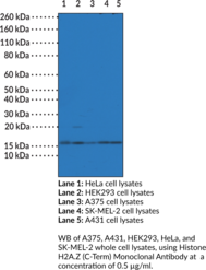
Histone H2A.Z is a variant of histone H2A, a nuclear protein and a component of the nucleosome core.{59522,57056} It is a globular protein containing unstructured N- and C-terminal tails that extend outside of the nucleosome core that are subject to a variety of post-translational modifications (PTMs), including acetylation, methylation, and ubiquitination, which function as epigenetic regulators of transcription.{58069} Histone H2A.Z influences chromatin accessibility and nucleosome stability, regulating various cellular functions, including DNA replication and repair, mitosis, heterochromatin formation, and chromosome segregation.{59522,59512} It is enriched at gene promoters and associated with active or repressed gene transcription in a manner dependent on species or PTMs, among other factors.{59526,59512} Increased tumor H2A.Z levels are associated with decreased overall survival in patients with breast cancer.{59527} Cayman’s Histone H2A.Z (C-Term) Monoclonal Antibody can be used for ELISA, immunocytochemistry (ICC), multiplex-based assay, and Western blot (WB) applications. The antibody recognizes the C-terminal region of histone H2A.Z independent of PTMs.
Brand:CaymanSKU:32184 - 100 µgAvailable on backorder
Histone H2A.Z is a highly conserved variant of the core histone H2A that comprises approximately 5% of the total H2A found in vertebrates.{60025} It is a globular protein that contains an unstructured N-terminal tail that extends outside of the nucleosome core and is subject to various post-translational modifications (PTMs), including methylation, phosphorylation, acetylation, SUMOylation and ubiquitination.{57056,60026} Acetylation of histone H2A.Z at lysine 4 (H2A.ZK4Ac) by histone acetyltransferase 5 (KAT5) is associated with transcriptional activation.{60026,60027} Cayman’s Histone H2A.ZK4Ac Monoclonal Antibody can be used for ELISA, immunocytochemistry (ICC), multiplex-based assay, and Western blot (WB) applications.
Brand:CaymanSKU:32171 - 100 µgAvailable on backorder
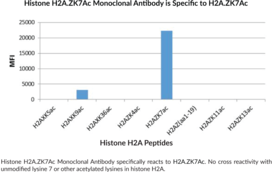
Histone H2A.Z is a highly conserved variant of the core histone H2A that comprises approximately 5% of the total H2A found in vertebrates.{60025} It is a globular protein that contains an unstructured N-terminal tail that extends outside of the nucleosome core and is subject to various post-translational modifications (PTMs), including methylation, phosphorylation, acetylation, SUMOylation and ubiquitination.{57056,60026} Acetylation of histone H2A.Z at lysine 7 (H2A.ZK7Ac) by the lysine acetyltransferase TIP60 is associated with transcriptional activation.{60026,60027} Cayman’s Histone H2A.ZK7Ac Monoclonal Antibody can be used for ELISA, immunocytochemistry (ICC), multiplex-based assay, and Western blot (WB) applications.
Brand:CaymanSKU:32172 - 100 µgAvailable on backorder
Histone H2AX is a variant of histone H2A, a nuclear protein and a component of the nucleosome core.{59522,57056} It is a globular protein containing unstructured N- and C-terminal tails that extend outside of the nucleosome core that are subject to a variety of post-translational modifications (PTMs), including phosphorylation, acetylation, methylation, and ubiquitination, which function as epigenetic regulators of transcription.{59523,58069} H2AX has a key role in the DNA damage response.{59523,58069,59522,59524} It is phosphorylated at serine 139 (γH2AX) by the PI3K-like kinases ATM, ATR, and DNA-PK in response to DNA damage, leading to changes in chromatin structure at the damaged site that promote DNA repair. H2AX has additional roles in chromatin inactivation during meiosis and mitosis, as well as neural stem cell development and cellular senescence.{59524} Decreased tumor H2AX levels are associated with increased progression-free survival in patients with triple-negative breast cancer.{59525} Cayman’s Histone H2AX (C-Term) Monoclonal Antibody can be used for ELISA, immunocytochemistry (ICC), multiplex-based assay, and Western blot (WB) applications. The antibody recognizes the C-terminal region of histone H2AX independent of PTMs.
Brand:CaymanSKU:32183 - 100 µgAvailable on backorder
