ELISA Kits
Showing 601–750 of 3623 results
-
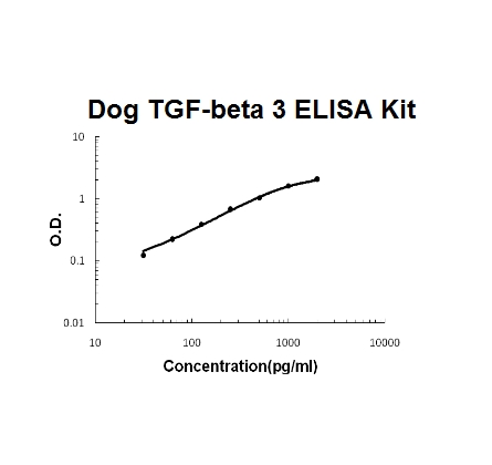
Sandwich High Sensitivity ELISA kit for Quantitative Detection of activated Dog Caninea TGF-beta 3. 96wells/kit, with removable strips.
Brand:Boster BioSKU:EK1103-CNAvailable on backorder
The dopamine transporter (DAT) is responsible for the reaccumulation of dopamine after it has been released. DAT antibodies and antibodies for other markers of catecholamine biosynthesis are widely used as markers for dopaminergic and noradrenergic neurons in a variety of applications including depression, schizophrenia, Parkinson’s disease, and drug abuse. Levels of DAT protein expression are altered by chronic drug administration.
Brand:CaymanSKU:10009372 - 1 eaAvailable on backorder
The dopamine transporter (DAT) is responsible for the reaccumulation of dopamine after it has been released. DAT antibodies and antibodies for other markers of catecholamine biosynthesis are widely used as markers for dopaminergic and noradrenergic neurons in a variety of applications including depression, schizophrenia, Parkinson’s disease, and drug abuse. Levels of DAT protein expression are altered by chronic drug administration.
Brand:CaymanSKU:10009373 - 1 eaAvailable on backorder
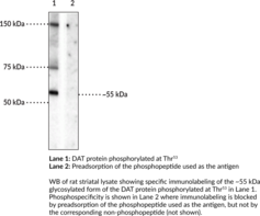
The dopamine transporter (DAT) is a member of the SLC6 family of transporters and is encoded by the SLC6A3 gene in humans.{55189,55190} DAT is expressed in dopaminergic neurons and localizes to perisynaptic sites, where it functions to translocate the neurotransmitter dopamine (DA; Item No. 21992) into presynaptic neurons from the extracellular space.{55191,55189} It is comprised of 12 transmembrane helices flanked by large cytoplasmic N- and C-terminal tails that are subject to protein-protein interactions with DAT binding proteins as well as posttranslational modifications.{55189,55191} DAT can be phosphorylated by ERK at threonine 53 (Thr53) in vitro.{55192} Phosphorylation of DAT at Thr53 (phospho-Thr53) is increased in rat striatal synaptosomes in response to phorbol 12-myristate 13-acetate (PMA; Item No. 10008014), okadaic acid (Item No. 10011490), amphetamine, or methamphetamine.{55192,55193} In vivo, phospho-Thr53 in rat striatum is increased following administration of methamphetamine.{55193} LLC-PK1 cells expressing rat DAT (rDAT) with non-phosphorylatable or phosphomimetic Thr53 mutations exhibit defects in DA uptake and amphetamine-induced efflux of the substrate [3H]MPP+ compared to cells expressing wild-type rDAT, indicating that phosphorylation of Thr53 has roles in regulating the uptake and efflux functions of DAT.{55192} Cayman’s Dopamine Transporter (Phospho-Thr53) Polyclonal Antibody can be used for Western blot (WB) applications. The antibody recognizes DAT (phospho-Thr53) at approximately 55 kDa from mouse and rat samples.
Brand:CaymanSKU:29261 - 100 µlAvailable on backorder
Dopamine β-hydroxylase (DBH) catalyzes the conversion of dopamine to norepinephrine and serves as a marker of noradrenergic cells. DBH antibodies and antibodies for other markers of catecholamine biosynthesis are widely used as markers for dopaminergic and noradrenergic neurons in a variety of applications including depression, schizophrenia, Parkinson’s disease, and drug abuse. The expression of DBH is also elevated during stress.
Brand:CaymanSKU:10009370 - 1 eaAvailable on backorder
Dopamine β-hydroxylase (DBH) catalyzes the conversion of dopamine to norepinephrine and serves as a marker of noradrenergic cells. DBH antibodies and antibodies for other markers of catecholamine biosynthesis are widely used as markers for dopaminergic and noradrenergic neurons in a variety of applications including depression, schizophrenia, Parkinson’s disease, and drug abuse. The expression of DBH is also elevated during stress.
Brand:CaymanSKU:10009371 - 1 eaAvailable on backorder
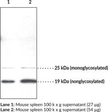
Doppel is a homolog of the cellular prion protein. Like prion protein, doppel has two N-linked oligosaccharides, and is presented on the cell surface via a glycosylphosphatidylinositol anchor.{11663} Unlike prion protein, it lacks the conformationally plastic and octapeptide repeat domains.{11604} The primary physiological role of doppel protein remains to be determined, but there is some evidence suggesting that cell surface prion protein can antagonize the toxic effect of doppel expressed in the central nervous system.{11663} In addition, the protein may play a major role in human male fertility, given its expression on both sertoli cells and spermatozoa.{11666} Doppel is differentially glycosylated, causing it to migrate at multiple sizes on SDS-PAGE.{11663} Cayman’s Doppel Polyclonal Antibody can be used for immunohistochemistry and Western blot applications. The antibody recognizes Doppel at 19 kDa (nonglycosylated) and 25 kDa (monoglycosylated) from human, mouse, and rat samples.
Brand:CaymanSKU:10005517 - 1 eaAvailable on backorder
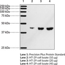
The DP1 receptor is one of two receptor isoforms for prostaglandin D2 (PGD2), a major cyclooxygenase metabolite of arachidonic acid. As a G-protein coupled receptor (GPCR), the receptor primarily couples to Gs to increase cAMP, but also transiently increases Ca2+, apparently through the cAMP pathway.{11488} DP1 receptor mRNA has been detected in various tissues and cells including brain, spleen, lung, bone marrow, stomach, skin, and mast cells,{3175, 3128,9114} however little is know about its protein localization due to its low level of expression. Cayman’s DP1 receptor polyclonal antibody detects the protein by western blot in samples such as cerebral cortex, hippocampus, and HT-29 cells, a colorectal cancer cell line shown to express this receptor.{12254}
Brand:CaymanSKU:101640 - 1 eaAvailable on backorder
Brand:CaymanSKU:700212 - 1 eaAvailable on backorder
DPP (IV) inhibitors have emerged as a new class of oral antidiabetic agents. These inhibitors promote glucose homeostasis by inhibiting degradation of glucose-dependent insulinotropic polypeptide (GIP) and glucagon-like peptide-1 (GLP-1) by DPP (IV). GLP-1 extends the action of insulin while suppressing the release of glucagon. Cayman’s DPP (IV) Inhibitor Screening Assay provides a convenient fluorescence-based method for screening DPP (IV) inhibitors in a 96-well format.
Brand:CaymanSKU:700210 - 96 wellsAvailable on backorder
Brand:CaymanSKU:700211 - 1 eaAvailable on backorder
Brand:CaymanSKU:700213 - 1 eaAvailable on backorder
In response to stimuli, neutrophils have the ability to release net-like structures containing nuclear DNA, de-condensed histones, and antimicrobial peptides.{19770} These neutrophil extracellular traps (NETs) have the ability to contact and kill pathogens including fungi, bacteria, and protozoa; they are then rapidly cleared by the immune system.{24590,24587,25672} However, in aged NZBWF1 mice and human lupus patients, the clearance is delayed, allowing formation of antibodies to these NET components. Cayman’s dsDNA Monoclonal Antibody was developed by fusing the spleen of a non-immunized NZBWF1 mouse with a mouse myeloma cell line. It detects dsDNA by ELISA and can be used to stain NETs by immunofluorescence.
Brand:CaymanSKU:15635 - 1 eaAvailable on backorder
Brand:CaymanSKU:760912 - 1 eaAvailable on backorder
Brand:CaymanSKU:700416 - 1 mlAvailable on backorder
Deubiquitinating enzymes (DUBs) remove ubiquitin from modified proteins in order to recycle ubiquitin attached to inappropriate targets, to remove and disassemble polyubiquitin chains, and to process proteins prior to their degradation by the proteasome.{26595} They have been implicated in a number of human diseases and thus, are attractive targets for potential therapeutic intervention via the development of suitable inhibitors and modulators.{26597,26595} Cayman’s DUB Activity Assay Kit facilitates the rapid, robust measurement of deubiquitinating enzyme activity in vitro. The kit utilizes a high purity, fluorogenic substrate (ubiquitin-AMC) together with suitable calibration standards and controls for the accurate and sensitive assessment of DUB activity. Continuous kinetic or end-point assays can be performed in a 96-well plate format for multi-sample analysis.
Brand:CaymanSKU:701490 - 96 wellsAvailable on backorder
The DYKDDDDK epitope is an expression tag commonly used in protein expression experiments. Compared to its corresponding native protein, a DYDDDDK-tagged protein will migrate at a larger size on SDS-PAGE, which is equivalent to the size of the epitope tag. The size of the DYDDDDK epitope may vary depending on the expression vector being utilized, but usually ranges from 3 to 5 kDa. Cayman Chemical’s DYDDDDK polyclonal antibody will recognize proteins expressed with both N-terminal and C-terminal DYDDDDK tags.
Brand:CaymanSKU:162150 - 1 eaAvailable on backorder
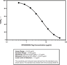
Cayman’s DYKDDDDK-Tag Detection ELISA Kit provides the ability to rapidly assess the levels of DYKDDDDK-tagged proteins at each stage of the expression and purification process. This permits the user to quickly monitor expression efforts and follow protein loss or enrichment at each purification step. This assay is designed for the rapid, semi-quantitative screening of cell lysates and affinity column fractions for DYKDDDDK-tagged proteins. It is intended to serve as a substitute for SDS-PAGE, thereby expediting the screening of affinity column fractions.
Brand:CaymanSKU:501560 - 96 wellsAvailable on backorder
Brand:CaymanSKU:10011231 - 1 eaAvailable on backorder
Dynamin is a member of a group of nerve terminal proteins called dephosphins that regulate synaptic vesicle endocytosis.{14791,14752,14751} Cyclin dependent protein kinase 5 phosphorylates dynamin at Ser774 and Ser778.{14792} Phosphorylation of these sites on dynamin is thought to play a key role in synaptic vesicle trafficking.
Brand:CaymanSKU:10009785 - 1 eaAvailable on backorder
Dynamin is a member of a group of nerve terminal proteins called dephosphins that regulate synaptic vesicle endocytosis.{14791,14752,14751} Cyclin dependent protein kinase 5 phosphorylates dynamin at Ser774 and Ser778. Phosphorylation of these sites on dynamin is thought to play a key role in synaptic vesicle trafficking.
Brand:CaymanSKU:10009786 - 1 eaAvailable on backorder
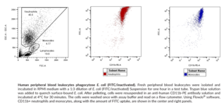
Cayman’s E. coli Phagocytosis Assay Kit employs FITC-labeled, heat-inactivated E. coli cells for the measurement of the phagocytic process in vitro. The engulfed fluorescent E. coli can be detected by flow cytometry.
Brand:CaymanSKU:601370 - 100 testsAvailable on backorder
Cayman’s Early Apoptosis Detection Assay Kit employs FITC-conjugated annexin V as a probe for phosphatidylserine (PS) on the outer membrane of apoptotic cells, TMRE as a probe for ΔΨm, DAPI as an indicator of membrane permeability/cell viability, and TO-PRO®-3 to indicate the selective permeability of pannexin channels. The Early Apoptosis Detection Assay Kit allows phenotypic characterization of multiple different cell death parameters at the single-cell level.
Brand:CaymanSKU:601360 - 100 testsAvailable on backorder
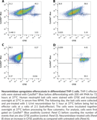
Efferocytosis is a specialized form of phagocytosis in which macrophages and other phagocytic cells clear dead and dying cells to promote homeostasis, embryonic development, regulation of the immune system and the resolution of inflammation.{35342} Efficient clearance of cells is essential for the removal of the nearly 1 million cells per second undergoing apoptosis in a human adult.{35343} Efferocytic removal of apoptotic cells before they become necrotic and release their contents can prevent an immune response to these non-pathogenic antigens.
Brand:CaymanSKU:601770 - 1 eaAvailable on backorder
Epidermal growth factor receptor (EGFR), also known as HER1 and ERBB1, is a cell surface receptor and member of the EGF family of receptor tyrosine kinases with roles in cell proliferation, differentiation, and survival.{54146,54144} It is a transmembrane receptor composed of an intracellular tyrosine kinase domain, a transmembrane lipophilic segment, and an extracellular domain that is expressed in epithelial, mesenchymal, and neuronal tissues.{54146,54144,54147} Under unstimulated conditions, EGFR is an auto-inhibited monomer in the plasma membrane.{54146} Upon canonical ligand binding, EGFR undergoes homodimerization or heterodimerization with HER2, HER3, or HER4, which induces a conformational change in the cytoplasmic domain that facilitates autophosphorylation and intracellular signaling. EGFR contains five C-terminal autophosphorylation sites, tyrosine 1068 (Tyr1068), Tyr1148, Tyr1173, Tyr1086, and Tyr992.{60064} EGFR autophosphorylation at Tyr1173 (phospho-Tyr1173) leads to interaction with phospholipase Cγ (PLCγ) and Shc, which have roles in activating the MAPK signaling pathway. Increased levels of EGFR (phospho-Tyr1173) are associated with poor progression-free survival in patients with non-small cell lung cancer (NSCLC). Levels of EGFR (phospho-Tyr1173) are increased in, and positively correlate with activation of MAPK signaling and cytokine production in, bronchial epithelial biopsies from healthy individuals exposed to diesel exhaust.{60065} Cayman’s EGFR (Phospho-Tyr1173) Rabbit Monoclonal Antibody can be used for immunohistochemistry (IHC) and Western blot (WB) applications.
Brand:CaymanSKU:32219 - 100 µlAvailable on backorder
This dye may be added to Cayman’s ELISA antisera if desired to aid in visualization of antiserum-containing wells.
Brand:CaymanSKU:400042 - 1 eaAvailable on backorder
Brand:CaymanSKU:400060 - 10 mlAvailable on backorder
Brand:CaymanSKU:400040 - 1 eaAvailable on backorder
Endothelial lipase (EL) is a major genetic determinant for the concentration, structure, and metabolism of high-density lipoprotein, which protects against atherosclerosis.{11500, 11501} It was originally cloned from endothelial cells and found to be expressed in a distinct and complementary tissue-restricted fashion, with high-level expression in the liver, placenta, lung, ovary, and macrophage.{11293} Immunohistochemical studies demonstrate that EL is expressed in infiltrating cells such as macrophages within atheromatous plaques, in addition to endothelial and smooth muscle cells in non-atherosclerotic coronary arteries. Furthermore, EL expression is detected in the neovasculature within atheromatous plaques in atherosclerotic coronary arteries, indicating that EL may have unique functional roles in atherosclerosis.{11499} Cayman Chemical’s EL polyclonal antibody can be used for western blot and immunohistochemical analysis for EL on samples of human, murine, rat, porcine, and ovine origin. Other applications for use of this antibody have not yet been tested.
Brand:CaymanSKU:100030 - 500 µlAvailable on backorder
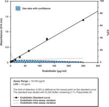
The endothelin peptide family consists of three isoforms, ET-1 (corresponding to the initially isolated and most predominant isoform), ET-2, and ET-3. ET-1 is a 21 amino acid peptide and is one of the most potent vasoconstritors currently known. ET-2 displays similar pharmacology to ET-1, whereas ET-3 is a weak vasoconstrictor but more potent inhibitor of platelet aggregation. Cayman’s Endothelin Assay is an immunometric (i.e., sandwich) ELISA that permits endothelin measurements within the range of 7.8-250 pg/ml, typically with a limit of detection of 7.8 pg/ml. Inter- and intra-assay CV’s of less than 10% may be achieved at most concentrations. This assay offers sensitive and specific analysis of endothelin in serum, plasma, urine, or cell culture media.
Brand:CaymanSKU:583151 - 480 wellsAvailable on backorder
The endothelin peptide family consists of three isoforms, ET-1 (corresponding to the initially isolated and most predominant isoform), ET-2, and ET-3. ET-1 is a 21 amino acid peptide and is one of the most potent vasoconstritors currently known. ET-2 displays similar pharmacology to ET-1, whereas ET-3 is a weak vasoconstrictor but more potent inhibitor of platelet aggregation. Cayman’s Endothelin Assay is an immunometric (i.e., sandwich) ELISA that permits endothelin measurements within the range of 7.8-250 pg/ml, typically with a limit of detection of 7.8 pg/ml. Inter- and intra-assay CV’s of less than 10% may be achieved at most concentrations. This assay offers sensitive and specific analysis of endothelin in serum, plasma, urine, or cell culture media.
Brand:CaymanSKU:583151 - 96 wellsAvailable on backorder
Brand:CaymanSKU:483154 - 1 eaAvailable on backorder
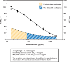
Enterolactone is a mammalian lignan with an estrogen-like diphenolic structure. It is produced by intestinal bacteria from two plant precursors (matairesinol and secoisolariciresinol) obtained in the diet. Enterolactone and other lignans and phytoestrogens have been associated with a reduced risk of acute coronary events, hormone-dependent cancers, and possibly osteoporosis. Several studies have suggested that serum enterolactone may serve as a biomarker of a healthy, high-fiber diet. Cayman Chemical’s Enterolactone ELISA kit is a competitive assay that utilizes a standard curve ranging from 15.6-2,000 pg/ml. The assay has a range from 15.6-2,500 pg/ml and a sensitivity (80% B/B0) of approximately 70 pg/ml.
Brand:CaymanSKU:500520 - 480 solid wellsAvailable on backorder
Enterolactone is a mammalian lignan with an estrogen-like diphenolic structure. It is produced by intestinal bacteria from two plant precursors (matairesinol and secoisolariciresinol) obtained in the diet. Enterolactone and other lignans and phytoestrogens have been associated with a reduced risk of acute coronary events, hormone-dependent cancers, and possibly osteoporosis. Several studies have suggested that serum enterolactone may serve as a biomarker of a healthy, high-fiber diet. Cayman Chemical’s Enterolactone ELISA kit is a competitive assay that utilizes a standard curve ranging from 15.6-2,000 pg/ml. The assay has a range from 15.6-2,500 pg/ml and a sensitivity (80% B/B0) of approximately 70 pg/ml.
Brand:CaymanSKU:500520 - 480 strip wellsAvailable on backorder
Enterolactone is a mammalian lignan with an estrogen-like diphenolic structure. It is produced by intestinal bacteria from two plant precursors (matairesinol and secoisolariciresinol) obtained in the diet. Enterolactone and other lignans and phytoestrogens have been associated with a reduced risk of acute coronary events, hormone-dependent cancers, and possibly osteoporosis. Several studies have suggested that serum enterolactone may serve as a biomarker of a healthy, high-fiber diet. Cayman Chemical’s Enterolactone ELISA kit is a competitive assay that utilizes a standard curve ranging from 15.6-2,000 pg/ml. The assay has a range from 15.6-2,500 pg/ml and a sensitivity (80% B/B0) of approximately 70 pg/ml.
Brand:CaymanSKU:500520 - 96 solid wellsAvailable on backorder
Enterolactone is a mammalian lignan with an estrogen-like diphenolic structure. It is produced by intestinal bacteria from two plant precursors (matairesinol and secoisolariciresinol) obtained in the diet. Enterolactone and other lignans and phytoestrogens have been associated with a reduced risk of acute coronary events, hormone-dependent cancers, and possibly osteoporosis. Several studies have suggested that serum enterolactone may serve as a biomarker of a healthy, high-fiber diet. Cayman Chemical’s Enterolactone ELISA kit is a competitive assay that utilizes a standard curve ranging from 15.6-2,000 pg/ml. The assay has a range from 15.6-2,500 pg/ml and a sensitivity (80% B/B0) of approximately 70 pg/ml.
Brand:CaymanSKU:500520 - 96 strip wellsAvailable on backorder
Brand:CaymanSKU:600341 - 20 µlAvailable on backorder
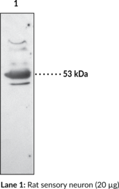
The biological effects of PGE2 are mediated through interaction with four distinct membrane-bound G-protein coupled EP receptors: EP1, EP2, EP3, and EP4.{1422,6742} Binding of PGE2 to the EP1 receptor results in an increase in phosphatidyl inositol turnover with subsequent increase in intracellular free Ca2+.{3176,2034} Pharmacologically, EP1 receptors mediate contraction of smooth muscle.{1422} The human EP1 receptor is comprised of 402 amino acids with a molecular mass of approximately 42,000.{3176} The EP1 receptor is expressed in a variety of tissues, including the kidney, lung, and sensory neuron.{3176,2034,9050} Within the kidney, the EP1 receptor is expressed at high levels in the cortical, outer medullary, and inner medullary collecting duct.{2773}
Brand:CaymanSKU:101740 - 1 eaAvailable on backorder
Prostaglandin E2 (PGE2) is one of the most important biologically active prostanoids which exerts its actions mainly by binding to four distinct E-type prostanoid receptors: EP1, EP2, EP3, and EP4. These four G protein-coupled receptors (GPCRs) exhibit differences in signal transduction mechanisms, tissue localization, and regulation of expression. EP2 receptors are expressed in many tissues and cells to mediate various PGE2 actions. The receptors couple to Gs to stimulate the cAMP second messenger signal transduction pathway. EP2 receptors play important roles in mucosal protection, gastrointestinal secretion, and motility. The diverse effects of PGE2 acting via EP2 receptors make it an interesting drug target. Therefore, EP2 subtype selective agonists, antagonists, and modulators have been identified with potential therapeutic applications.
Brand:CaymanSKU:601380 - 96 wellsAvailable on backorder
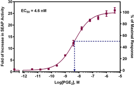
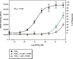
The EP2 receptor is one of four GPCRs that mediate the actions of PGE2. The diverse effects of PGE2 acting via EP2 receptors point to the need to identify novel agonists and antagonists, both to further elucidate the function of this receptor subtype and for use as therapeutics for various diseases. Cayman’s EP2 Receptor (rat) Reporter Assay Kit consists of a 96-well plate coated with a transfection complex containing DNA constructs for rat EP2 receptor and a cAMP response element regulated Secreted Alkaline Phosphatase (SEAP) reporter (EP2 Receptor Reverse Transfection Plate). Cells grown on the transfection complex will express EP2 at the cell surface. Binding of agonists to EP2 initiates a signal transduction cascade resulting in expression of SEAP which is secreted into the cell culture media. Aliquots of media are removed at time intervals, beginning at approximately six hours, and SEAP activity is measured following addition of a luminescence-based alkaline phosphatase substrate provided in the kit. The kit is simple to use and can be easily adapted to high throughput screening for therapeutic compounds regulating the activation of EP2. PGE2 is included in the kit for use as a positive control. The kit provides sufficient reagents to measure SEAP activity at three time points using the black plates included.
Brand:CaymanSKU:600340 - 100 testsAvailable on backorder
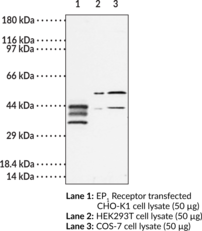
The biological effects of prostaglandin E2 (PGE2) are mediated through interaction with four distinct membrane-bound G-protein coupled EP receptors: EP1, EP2, EP3, and EP4.{1422,6741} Binding of PGE2 to the EP2 receptor results in an increase in adenylate cyclase activity with a subsequent increase in cAMP.{3178,5500} Pharmacologicially, EP2 receptors mediate relaxation of smooth muscle and are distinguished from EP4 receptors by their sensitivity to the EP2 receptor selective agonist butaprost.{1422,6741,3178} The human EP2 receptor is comprised of 358 amino acids with a molecular mass of approximately 40,000.{3178} EP2 is detected on immunoblot at 65 or 52 kDa depending on the degree of post-translational modifications of that receptor. mRNA for the EP2 receptor is expressed in a variety of tissues including lung, placenta, spleen, intestine, kidney, and sensory neuron.{3178,5500,9050}
Brand:CaymanSKU:101750 - 500 µlAvailable on backorder
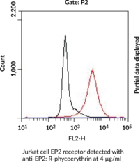
This polyclonal antibody is labeled with R-Phycoerythrin (PE) and can be used for immunofluorescent labeling of cellular EP2 receptor following permeabilization. R-PE absorption maxima occurs at 565>540>498 nm and the emission maximum occurs at 578 nm, however standard excitation by 488 nm lasers is sufficient to generate signal detected in the PE channel of flow cytometers. The biological effects of prostaglandin E2 (PGE2) are mediated through interaction with four distinct membrane-bound G-protein coupled EP receptors: EP1, EP2, EP3, and EP4.{1422,4345} Binding of PGE2 to the EP2 receptor results in an increase in adenylate cyclase activity with a subsequent increase in cAMP.{10096,5500} Pharmacologically, EP2 receptors mediate relaxation of smooth muscle and are distinguished from EP4 receptors by their sensitivity to the EP2 receptor selective agonist butaprost.{1422,4345,10096} The human EP2 receptor is comprised of 358 amino acids with a molecular mass of approximately 40,000.{10096} mRNA for the EP2 receptor is expressed in a variety of tissues including lung, placenta, spleen, intestine, kidney, and sensory neuron.{10096,5500,9050}
Brand:CaymanSKU:10477 - 1 eaAvailable on backorder
The biological effects of prostaglandin E2 (PGE2) are mediated through interaction with four distinct membrane-bound G-protein coupled EP receptors: EP1, EP2, EP3, and EP4.{6742,9114} As a result of splice variation, the EP3 receptor can be expressed as multiple isoforms that differ in the length and sequence of their C-terminal tails.{9114,3180} The signal transduction mechanism varies depending on the isoform being expressed, implicating the importance of the C-terminal region of the receptor for coupling to G-proteins.{9114,3180} The EP3 receptor is expressed in a variety of tissues with highest levels in kidney, pancreas, and uterus.{3180,1610,3179}
Brand:CaymanSKU:101760 - 1 eaAvailable on backorder
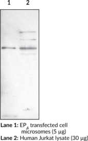
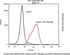
The biological effects of prostaglandin E2 (PGE2) are mediated through interaction with four distinct membrane-bound G-protein coupled EP receptors: EP1, EP2, EP3, and EP4.{6742,9114} Binding of PGE2 to the EP4 receptor causes an increase in intracellular cyclic AMP and subsequent relaxation of smooth muscle.{3164,3186,2035,3185} In addition to sequence differences, EP4 receptors are distinguished from EP2 receptors by their insensitivity to the EP2 receptor selective agonist butaprost.{2035,3178} The EP4 receptor is expressed in multiple human tissues including kidney, small intestine, thymus, ileum, uterus, and lung, as well as in peripheral blood leukocytes.{3164,3186} The receptor is also expressed in cultured human blood cell lines of monocytoid (U937 and THP-1) and lymphoid (MOLT-4, Jurkat, and Raji) origin.{3185}
Brand:CaymanSKU:101775 - 1 eaAvailable on backorder
The biological effects of prostaglandin E2 (PGE2) are mediated through interaction with four distinct membrane-bound G protein-coupled EP receptors: EP1, EP2, EP3, and EP4.{6742,9114} Binding of PGE2 to the EP4 receptor causes an increase in intracellular cyclic AMP and subsequent relaxation of smooth muscle.{3164,3186,2035,3185} In addition to sequence differences, EP4 receptors are distinguished from EP2 receptors by their insensitivity to the EP2 receptor selective agonist butaprost.{2035,3178} The EP4 receptor is expressed in multiple human tissues including kidney, small intestine, thymus, ileum, uterus, and lung, as well as in peripheral blood leukocytes.{3164,3186} The receptor is also expressed in cultured human blood cell lines of monocytoid (U937 and THP-1) and lymphoid (MOLT-4, Jurkat, and Raji) origin.{3185}
Brand:CaymanSKU:10479 - 500 µlAvailable on backorder
Prostaglandin E2 (PGE2) is one of the most important biologically active prostanoids which exerts its actions mainly by binding to four distinct E-type prostanoid receptors: EP1, EP2, EP3, and EP4. These four G protein-coupled receptors (GPCRs) exhibit differences in signal transduction mechanisms, tissue localization, and regulation of expression. EP4 receptors are highly expressed in the intestine, but are found in lower levels in the lung, kidney, thymus, uterus, and brain. The receptors are primarily coupled to Gs to stimulate the cAMP second messenger signal transduction pathway. EP4 receptors play important roles in relaxation of smooth muscle, gastric acid secretion, intestinal epithelial transportation, adrenal aldosterone secretion, uterine functions, bone formation, bone resorption, immunity, and inflammation. They have been suggested to involve in colorectal carcinogenesis, neuroprotection in excitotoxic brain injury, and cardiac hypertrophy. The diverse effects of PGE2 via EP4 receptors point to the need to identify novel agonists, antagonists, and modulators, both to further elucidate the function of this receptor subtype and for use as therapeutics for various diseases.
Brand:CaymanSKU:601390 - 96 wellsAvailable on backorder
The EP4 receptor is one of four GPCRs that mediate the actions of prostaglandin E2 (PGE2). The diverse effects of PGE2 acting via EP4 receptors point to the need to identify novel agonists and antagonists, both to further elucidate the function of this receptor subtype and for use as therapeutics for various diseases. Cayman’s EP4 Receptor (rat) Activation Assay Kit (cAMP) consists of a 96-well plate coated with a transfection complex containing a DNA construct for rat EP4 Receptor (EP4 Reverse Transfection Strip Plate). Cells grown on the transfection complex will express EP4 at the cell surface. Binding of agonists to EP4 stimulates cAMP generation and increases intracellular cAMP levels, which can be measured by a competitive EIA using the Mouse Anti-Rabbit IgG Coated Plate and the reagents included in the kit. An EP4 agonist, PGE2, is included in the kit as a positive control.
Brand:CaymanSKU:600410 - 1 eaAvailable on backorder
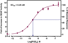
The EP4 receptor is one of four GPCRs that mediate the actions of prostaglandin E2 (PGE2). The diverse effects of PGE2 acting via EP4 receptors point to the need to identify novel agonists and antagonists, both to further elucidate the function of this receptor subtype and for use as therapeutics for various diseases. Cayman’s EP4 Receptor (rat) Reporter Assay Kit consists of a 96-well plate coated with a transfection complex containing DNA constructs for rat EP4 receptor and a cAMP response element regulated Secreted Alkaline Phosphatase (SEAP) reporter (EP4 Receptor Reverse Transfection Strip Plate). Cells grown on the transfection complex will express EP4 at the cell surface. Binding of agonists to EP4 initiates a signal transduction cascade resulting in expression of SEAP which is secreted into the cell culture media. Aliquots of media are removed at time intervals, beginning at approximately six hours, and SEAP activity is measured following addition of a luminescence-based alkaline phosphatase substrate provided in the kit. The kit is simple to use and can be easily adapted to high throughput screening for therapeutic compounds regulating the activation of EP4. PGE2 is included in the kit for use as a positive control. The kit provides sufficient reagents to measure SEAP activity at three time points using the black plates included.
Brand:CaymanSKU:600350 - 100 testsAvailable on backorder
Extracellular-signal regulated kinase/mitogen-activated protein kinase (ERK/MAPK) is an integral component of cellular signaling during mitogenesis and differentiation of mitotic cells and also is thought to play a key role in learning and memory.{14317,14327,14328,14333} The activity of this kinase is regulated by dual phosphorylation at Thr202 and Tyr204.{14328}
Brand:CaymanSKU:10009179 - 1 eaAvailable on backorder
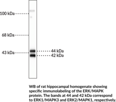
Extracellular-signal-regulated kinase/mitogen-activated protein kinases (ERK/MAPKs) are a widely conserved family of serine/threonine protein kinases and essential components of the MAP kinase signaling cascade.{53684,53685} Upon extracellular stimulation with mitogens, growth factors, or cytokines, a sequential three-part signaling cascade is initiated that terminates with phosphorylation of ERK/MAPK by MEK1 or MEK2. Activated ERK/MAPK is translocated to the nucleus where it regulates transcription factor activity and gene transcription for a variety of cellular processes including cell proliferation, differentiation, and stress response. Misregulation of the MAP kinase cascade drives development of various cancers. ERK1/MAPK3 and ERK2/MAPK1 are two key members of the ERK/MAPK family encoded by MAPK3 and MAPK1, respectively, in humans and are expressed in all tissues. Cayman’s ERK/MAPK Polyclonal Antibody can be used for immunocytochemistry (ICC) and Western blot (WB) applications. The antibody recognizes ERK1/MAPK3 and ERK2/MAPK1 at approximately 44 and 42 kDa, respectively from human, mouse, and rat samples.
Brand:CaymanSKU:29263 - 100 µlAvailable on backorder
Brand:CaymanSKU:501891 - 100 dtnAvailable on backorder
Brand:CaymanSKU:501891 - 500 dtnAvailable on backorder
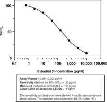
Estradiol is a steroid hormone produced from testosterone via the aromatase system in the granulosa cells of ovarian follicles.{42700,8343} It is instrumental in the development of secondary sex characteristics at puberty and in the menstrual cycle.{42701,42702,8458} Plasma levels of estradiol peak during the follicular phase of the menstrual cycle to approximately 300 pg/ml.{42700,42702,8458} During this time, it stimulates proliferation of granulosa cells, increases the size of uterine glands, and exerts positive feedback on the hypothalamus, leading to an increase in luteinizing hormone.{8458,42704} Blood levels of estradiol drop as luteinizing hormone levels increase and trigger ovulation, then rise again during the luteal phase to approximately 100 pg/ml.{42702} Estradiol is metabolized into estrone by 17β-hydroxysteroid dehydrogenase 2 and hydroxylated metabolites such as estriol, as well as glucuronidated and sulfonated metabolites, which are excreted in the urine and feces.{42703}
Brand:CaymanSKU:501890 - 480 solid wellsAvailable on backorder
Estradiol is a steroid hormone produced from testosterone via the aromatase system in the granulosa cells of ovarian follicles.{42700,8343} It is instrumental in the development of secondary sex characteristics at puberty and in the menstrual cycle.{42701,42702,8458} Plasma levels of estradiol peak during the follicular phase of the menstrual cycle to approximately 300 pg/ml.{42700,42702,8458} During this time, it stimulates proliferation of granulosa cells, increases the size of uterine glands, and exerts positive feedback on the hypothalamus, leading to an increase in luteinizing hormone.{8458,42704} Blood levels of estradiol drop as luteinizing hormone levels increase and trigger ovulation, then rise again during the luteal phase to approximately 100 pg/ml.{42702} Estradiol is metabolized into estrone by 17β-hydroxysteroid dehydrogenase 2 and hydroxylated metabolites such as estriol, as well as glucuronidated and sulfonated metabolites, which are excreted in the urine and feces.{42703}
Brand:CaymanSKU:501890 - 480 strip wellsAvailable on backorder
Estradiol is a steroid hormone produced from testosterone via the aromatase system in the granulosa cells of ovarian follicles.{42700,8343} It is instrumental in the development of secondary sex characteristics at puberty and in the menstrual cycle.{42701,42702,8458} Plasma levels of estradiol peak during the follicular phase of the menstrual cycle to approximately 300 pg/ml.{42700,42702,8458} During this time, it stimulates proliferation of granulosa cells, increases the size of uterine glands, and exerts positive feedback on the hypothalamus, leading to an increase in luteinizing hormone.{8458,42704} Blood levels of estradiol drop as luteinizing hormone levels increase and trigger ovulation, then rise again during the luteal phase to approximately 100 pg/ml.{42702} Estradiol is metabolized into estrone by 17β-hydroxysteroid dehydrogenase 2 and hydroxylated metabolites such as estriol, as well as glucuronidated and sulfonated metabolites, which are excreted in the urine and feces.{42703}
Brand:CaymanSKU:501890 - 96 solid wellsAvailable on backorder
Estradiol is a steroid hormone produced from testosterone via the aromatase system in the granulosa cells of ovarian follicles.{42700,8343} It is instrumental in the development of secondary sex characteristics at puberty and in the menstrual cycle.{42701,42702,8458} Plasma levels of estradiol peak during the follicular phase of the menstrual cycle to approximately 300 pg/ml.{42700,42702,8458} During this time, it stimulates proliferation of granulosa cells, increases the size of uterine glands, and exerts positive feedback on the hypothalamus, leading to an increase in luteinizing hormone.{8458,42704} Blood levels of estradiol drop as luteinizing hormone levels increase and trigger ovulation, then rise again during the luteal phase to approximately 100 pg/ml.{42702} Estradiol is metabolized into estrone by 17β-hydroxysteroid dehydrogenase 2 and hydroxylated metabolites such as estriol, as well as glucuronidated and sulfonated metabolites, which are excreted in the urine and feces.{42703}
Brand:CaymanSKU:501890 - 96 strip wellsAvailable on backorder
Brand:CaymanSKU:482282 - 100 dtnAvailable on backorder
Brand:CaymanSKU:482282 - 500 dtnAvailable on backorder
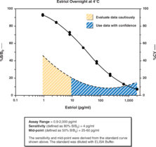
Estriol, a metabolite of 17β-estradiol and a major estrogen produced during pregnancy, has historically been used as an indicator of fetal well-being. In the final weeks before parturition, estriol levels increase significantly. In a longitudinal study in healthy pregnant women, total plasma estriol levels increased from 10 weeks gestation to approximately 150 ng/ml at week 38.{7767} The majority of the estriol synthesized in the later stages of pregnancy originates from fetal dehydroepiandrosterone sulfate (DHEAS) and serves as a direct marker of fetal adrenal gland activity.{7766} In the 1960s and 1970s plasma and urine were used to measure estriol. However, saliva contains primarily unbound and unconjugated estriol, the biologically active form of the hormone, and therefore is currently a commonly used biological fluid for monitoring estriol levels.{7766,7799} Plasma levels of estriol in males and non-pregnant females is less than 2 ng/ml.{7647}
Brand:CaymanSKU:582281 - 480 solid wellsAvailable on backorder
Estriol, a metabolite of 17β-estradiol and a major estrogen produced during pregnancy, has historically been used as an indicator of fetal well-being. In the final weeks before parturition, estriol levels increase significantly. In a longitudinal study in healthy pregnant women, total plasma estriol levels increased from 10 weeks gestation to approximately 150 ng/ml at week 38.{7767} The majority of the estriol synthesized in the later stages of pregnancy originates from fetal dehydroepiandrosterone sulfate (DHEAS) and serves as a direct marker of fetal adrenal gland activity.{7766} In the 1960s and 1970s plasma and urine were used to measure estriol. However, saliva contains primarily unbound and unconjugated estriol, the biologically active form of the hormone, and therefore is currently a commonly used biological fluid for monitoring estriol levels.{7766,7799} Plasma levels of estriol in males and non-pregnant females is less than 2 ng/ml.{7647}
Brand:CaymanSKU:582281 - 480 strip wellsAvailable on backorder
Estriol, a metabolite of 17β-estradiol and a major estrogen produced during pregnancy, has historically been used as an indicator of fetal well-being. In the final weeks before parturition, estriol levels increase significantly. In a longitudinal study in healthy pregnant women, total plasma estriol levels increased from 10 weeks gestation to approximately 150 ng/ml at week 38.{7767} The majority of the estriol synthesized in the later stages of pregnancy originates from fetal dehydroepiandrosterone sulfate (DHEAS) and serves as a direct marker of fetal adrenal gland activity.{7766} In the 1960s and 1970s plasma and urine were used to measure estriol. However, saliva contains primarily unbound and unconjugated estriol, the biologically active form of the hormone, and therefore is currently a commonly used biological fluid for monitoring estriol levels.{7766,7799} Plasma levels of estriol in males and non-pregnant females is less than 2 ng/ml.{7647}
Brand:CaymanSKU:582281 - 96 solid wellsAvailable on backorder
Estriol, a metabolite of 17β-estradiol and a major estrogen produced during pregnancy, has historically been used as an indicator of fetal well-being. In the final weeks before parturition, estriol levels increase significantly. In a longitudinal study in healthy pregnant women, total plasma estriol levels increased from 10 weeks gestation to approximately 150 ng/ml at week 38.{7767} The majority of the estriol synthesized in the later stages of pregnancy originates from fetal dehydroepiandrosterone sulfate (DHEAS) and serves as a direct marker of fetal adrenal gland activity.{7766} In the 1960s and 1970s plasma and urine were used to measure estriol. However, saliva contains primarily unbound and unconjugated estriol, the biologically active form of the hormone, and therefore is currently a commonly used biological fluid for monitoring estriol levels.{7766,7799} Plasma levels of estriol in males and non-pregnant females is less than 2 ng/ml.{7647}
Brand:CaymanSKU:582281 - 96 strip wellsAvailable on backorder
Brand:CaymanSKU:482284 - 1 eaAvailable on backorder
There are two classes of estrogen receptors (ERs). The first is intracellular and translocate to the nucleus when activated through binding to 17 beta estradiol. They are named ER alpha and ER beta. The second G protein couples receptors which are membrane proteins and have intracellular signaling function. Central ERα signaling is necessary for the actions of GLP-1 on food-reward behavior. [Bertin Catalog No. G01008]
Brand:CaymanSKU:32766 - 100 µlAvailable on backorder
There are two classes of Estrogen Receptors (ER,) the first is intracellular and translocate to the Nucleus when activated through binding to 17 beta estradiol. They are named ER alpha and ER beta. The second G protein couples receptors which are membranes proteins and have intracellular signaling function. central ERα signaling is necessary for the actions of GLP-1 on food-reward behavior. [Bertin Catalog No. G01009]
Brand:CaymanSKU:32767 - 100 µlAvailable on backorder
There are two classes of Estrogen Receptors (ERs) the first is intracellular and translocate to the nucleus when activated through binding to 17 beta estradiol. They are named ER alpha and ER beta. The second G protein couples receptors which are membranes proteins and have intracellular signaling function. Central ERα signaling is necessary for the actions of GLP-1 on food-reward behavior. [Bertin Catalog No. G01007]
Brand:CaymanSKU:32765 - 100 µlAvailable on backorder
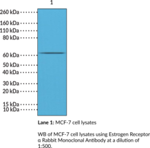
Estrogen receptor α (ERα) is a member of the nuclear receptor superfamily of transcription factors encoded by ESR1 in humans that has a key role in reproductive function and additional roles in cancer progression and inhibition.{59658,59659,59660} Alternative splicing of the ESR1 pre-mRNA produces one full-length 66 kDa isoform and two short-length 46 and 36 kDa isoforms, which contain truncated C-termini.{59660} ERα is comprised of an N-terminal domain that contains AF-1, which is critical for the transactivation function of ERα, a DNA binding domain that recognizes estrogen response elements (EREs) on target genes, and a C-terminal ligand binding domain (LBD) that contains the nuclear localization signal.{59659,59658} ERα is widely expressed in numerous tissues, including breast, prostate, uterus, liver, and bone, and localizes to the cytoplasm in a complex with heat shock protein 90 (Hsp90).{59660,59659} Upon estrogen stimulation, ERα dissociates from Hsp90, dimerizes, and translocates to the nucleus, where it binds EREs expressed by target genes encoding proteins that activate numerous signaling pathways, including ERK/MAPK, PI3K/Akt/mTOR, and NF-κB, and influence a variety of cellular processes, including reproductive function, bone homeostasis, and inflammation, as well as tumor progression and metastasis.{59659,59658} ERα is overexpressed in more than 50% of patients with breast cancer, but is associated with improved prognosis, whereas loss of ERα is a feature of triple-negative breast cancer (TNBC) and is associated with resistance to hormonal therapy.{59661} Cayman’s Estrogen Receptor α Rabbit Monoclonal Antibody can be used for immunohistochemistry (IHC) and Western blot (WB) applications.
Brand:CaymanSKU:32234 - 100 µlAvailable on backorder
There are two classes of estrogen receptors (ERs). The first is intracellular and translocate to the Nucleus when activated through binding to 17 beta estradiol. They are named ER alpha and ER beta. The second G protein couples receptors which are membrane proteins and have intracellular signaling function. Central ERα signaling is necessary for the actions of GLP-1 on food-reward behavior. [Bertin Catalog No. G01010]
Brand:CaymanSKU:32768 - 100 µlAvailable on backorder
There are two classes of estrogen receptors (ER,) the first is intracellular and translocate to the nucleus when activated through binding to 17β estradiol. They are named ERα and ERβ. The second G protein couples receptors which are membranes proteins and have intracellular signaling function. Central ERα signaling is necessary for the actions of GLP-1 on food-reward behavior. [Bertin Catalog No. G01011]
Brand:CaymanSKU:32769 - 100 µlAvailable on backorder
There are two classes of Estrogen Receptors (ER), the first is intracellular and translocate to the nucleus when activated through binding to 17β estradiol. They are named ERα and ERβ. The second G protein couples receptors which are membranes proteins and have intracellular signaling function. Central ERα signaling is necessary for the actions of GLP-1 on food-reward behavior. [Bertin Catalog No. G01012]
Brand:CaymanSKU:32770 - 100 µLAvailable on backorder
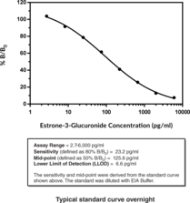
Estrone-3-glucuronide is the predominant metabolite of estradiol in urine and a urinary marker for the fertile window in women.{40240} Measurement of urinary glucuronides is a convenient, non-invasive method for detecting reproductive hormone levels compared to measuring plasma levels.{40241} Estrone-3-glucuronide, along with pregnanediol-3-glucuronide, are indicators of female reproductive health.{40241} Literature has shown high serum estrone levels, which directly correlate to urinary estrone-3-glucuronide levels, are associated with estrogen receptor-positive breast cancers.{40244} Hormone receptor status is the main factor in planning treatment and can be treated with hormone therapies, including tamoxifen and aromatase inhibitors.{40247} In addition to its role in breast cancer, circulating estrogens, including estrone metabolized from oral hormone replacement therapy, have been shown to increase thrombin generation leading to higher risk for blood clots, myocardial infarctions, deep vein thrombosis and strokes.{40245,40246}
Brand:CaymanSKU:501290 - 96 solid plateAvailable on backorder
Estrone-3-glucuronide is the predominant metabolite of estradiol in urine and a urinary marker for the fertile window in women.{40240} Measurement of urinary glucuronides is a convenient, non-invasive method for detecting reproductive hormone levels compared to measuring plasma levels.{40241} Estrone-3-glucuronide, along with pregnanediol-3-glucuronide, are indicators of female reproductive health.{40241} Literature has shown high serum estrone levels, which directly correlate to urinary estrone-3-glucuronide levels, are associated with estrogen receptor-positive breast cancers.{40244} Hormone receptor status is the main factor in planning treatment and can be treated with hormone therapies, including tamoxifen and aromatase inhibitors.{40247} In addition to its role in breast cancer, circulating estrogens, including estrone metabolized from oral hormone replacement therapy, have been shown to increase thrombin generation leading to higher risk for blood clots, myocardial infarctions, deep vein thrombosis and strokes.{40245,40246}
Brand:CaymanSKU:501290 - 96 strip plateAvailable on backorder
Brand:CaymanSKU:700566 - 2 mlAvailable on backorder
ETS is one of the largest family of TF (with 28 members in Human). Protein C-ets-1 is a protein that in humans is encoded by the ETS1. ETS1 is playing an important role in immune cells, and tissues. [Bertin Catalog No. G01013]
Brand:CaymanSKU:32771 - 100 µlAvailable on backorder
ETS is one of the largest family of TF (with 28 members in Human). Protein C-ets-1 is a protein that in humans is encoded by the ETS1. ETS1 is playing an important role in immune cells and tissues. [Bertin Catalog No. G01014]
Brand:CaymanSKU:32772 - 100 µlAvailable on backorder
Brand:CaymanSKU:700301 - 1 eaAvailable on backorder
Brand:CaymanSKU:10010317 - 1 eaAvailable on backorder
Brand:CaymanSKU:10010376 - 1 eaAvailable on backorder
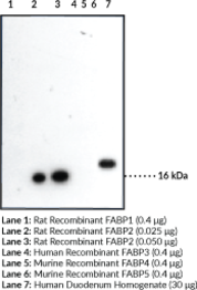
Fatty acid binding protein 2 (FABP2) is one of nine known cytosolic FABPs ranging in size from 14-15 kDa containing 127-133 amino acids.{11979} Members of this protein family exhibit high affinity for small lipophilic ligands and were named according to the tissue from which they were initially isolated. Studies suggest that FABPs are involved in the uptake and metabolism of fatty acids, in the maintenance of cellular membrane fatty acid levels, in intracellular trafficking of these substrates, in the modulation of specific enzymes of lipid metabolic pathways, and in the regulation of cell growth and differentiation.{11977} FABP family members have highly conserved three dimensional structures and 22-73% amino acid sequence similarity. FABP2 is composed of ten antiparallel β strands that form a barrel that binds ligand in a bent conformation. FABP2 polymorphism has been suggested to be associated with gender specific obesity and increased risk of diabetes.{11979} Cayman’s FABP2 Polyclonal Antibody can be used for western blot applications. The antibody recognizes FABP2 at 15.207 kDa from human and rat samples.
Brand:CaymanSKU:10010019 - 500 µlAvailable on backorder
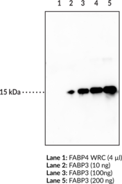
Fatty acid binding protein 3 (FABP3) is one of nine known cytosolic FABPs ranging in size from 14-15 kDa containing 127-132 amino acids.{11979} Members of this protein family exhibit high affinity for small lipophilic ligands and were named according to the tissue from which they were initially isolated.{11979} Studies suggest that FABPs are involved in the uptake and metabolism of fatty acids, in the maintenance of cellular membrane fatty acid levels, in intracellular trafficking of these substrates, in the modulation of specific enzymes of lipid metabolic pathways, and in the modulation of cell growth and differentiation.{11977} FABP family members have highly conserved three dimensional structures and 22-73% amino acid sequence similarity. FABP3 is composed of ten antiparallel β strands that form a barrel and is the most widely distributed FABP. It is found in heart, skeletal and smooth muscle, mammary epithelial cells, aorta, distal tubules of the kidney, lung, brain, placenta, and ovary. FABP3 is a potential biomarker for myocardial injury, especially for early detection of acute myocardial infarction (AMI).{11979}
Brand:CaymanSKU:10233 - 1 eaAvailable on backorder
Brand:CaymanSKU:10010318 - 1 eaAvailable on backorder
