ELISA Kits

Showing 2551–2700 of 3623 results

  • For quantitative detection of mouse SLIT2 in cell culture supernates, serum and plasma (heparin, EDTA).   


    Brand:
    Boster Bio
    SKU:EK1860

    Available on backorder
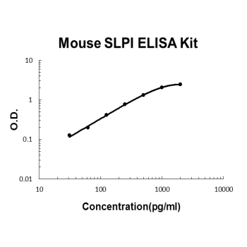
  • For quantitative detection of mouse SLPI in cell culture supernates, serum and plasma (heparin, EDTA).   


    Brand:
    Boster Bio
    SKU:EK1996

    Available on backorder
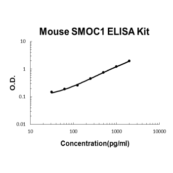
  • For quantitative detection of mouse SMOC1 in cell culture supernates, serum and plasma (heparin).   


    Brand:
    Boster Bio
    SKU:EK1936

    Available on backorder
  • For quantitative detection of mouse SP-D in cell culture supernates, cell lysates, serum and plasma (heparin, EDTA).   


    Brand:
    Boster Bio
    SKU:EK1186

    Available on backorder
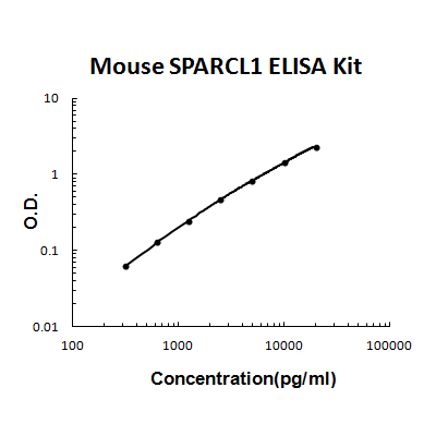
  • For quantitative detection of mouse SPARCL1 in cell culture supernates, serum and plasma (heparin, EDTA, citrate).   


    Brand:
    Boster Bio
    SKU:EK2125

    Available on backorder
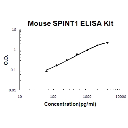
  • For quantitative detection of mouse SPINT1 in cell culture supernates, serum and plasma(heparin).   


    Brand:
    Boster Bio
    SKU:EK0772

    Available on backorder
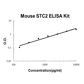
  • For quantitative detection of mouse STC2 in cell culture supernates, serum and plasma (heparin).   


    Brand:
    Boster Bio
    SKU:EK1989

    Available on backorder
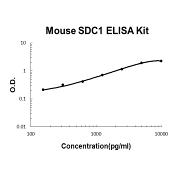
  • For quantitative detection of mouse Syndecan-1 in cell culture supernates , cell lysates, serum and plasma(heparin, EDTA).   


    Brand:
    Boster Bio
    SKU:EK1554

    Available on backorder
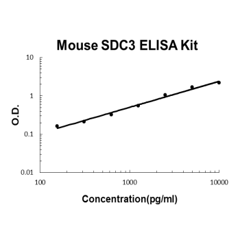
  • For quantitative detection of mouse Syndecan-3 in cell culture supernates , cell lysates, serum and plasma(heparin, EDTA).   


    Brand:
    Boster Bio
    SKU:EK1556

    Available on backorder
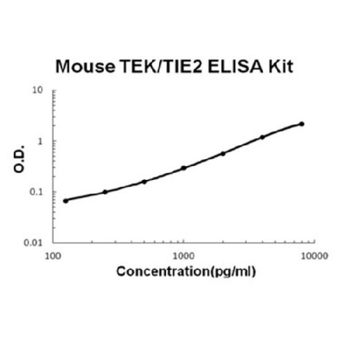
  • For quantitative detection of mouse TEK in cell culture supernates, serum and plasma(heparin, EDTA).   


    Brand:
    Boster Bio
    SKU:EK0976

    Available on backorder
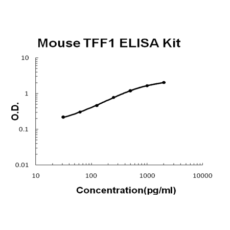
  • For Quantitative Detection of mouse TFF1 in cell culture supernates, serum and plasma (heparin, EDTA).   


    Brand:
    Boster Bio
    SKU:EK1620

    Available on backorder
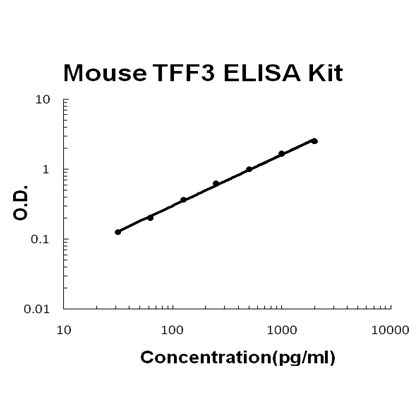
  • For Quantitative Detection of mouse TFF3 in cell culture supernates, serum and plasma (heparin, EDTA).   


    Brand:
    Boster Bio
    SKU:EK1629

    Available on backorder
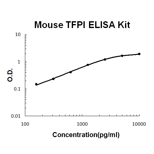
  • For quantitative detection of mouse TFPI in cell culture supernates, cell lysates, serum and plasma (heparin, EDTA).   


    Brand:
    Boster Bio
    SKU:EK1355

    Available on backorder
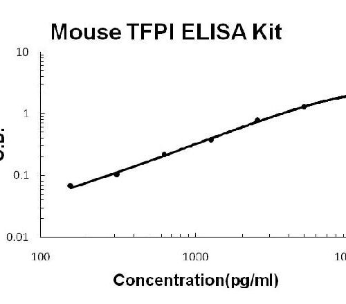
  • The Fast version of Picokine ELISA kits, assay takes less than 1.5 hours. Detect Mouse TFPI with <10pg/ml sensitivity. Format: 96-well plate with removable strips. Compatible samples: cell culture supernates, cell lysates, serum and plasma (heparin, EDTA). This is a TMB colorimetric sandwich ELISA kit with short assay time and fast experiment set up. TFPI tissue specificity: Isoform alpha is expressed in heart and spleen; isoform beta in heart and lung.   


    Brand:
    Boster Bio
    SKU:FEK1355

    Available on backorder
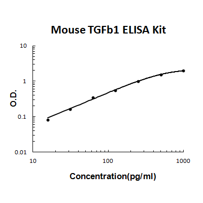
  • For quantitative detection of activated mouse TGF beta1 in cell culture supernates, serum, plasma(EDTA) and urine.   


    Brand:
    Boster Bio
    SKU:EK0515

    Available on backorder
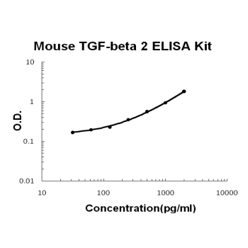
  • For quantitative detection of activated mouse TGF-beta 2 in cell culture supernates, serum and plasma(heparin, EDTA, citrate).   


    Brand:
    Boster Bio
    SKU:EK0983

    Available on backorder
  • For quantitative detection of activated mouse TGF-beta 3 in cell culture supernates, serum, plasma(EDTA) and urine.   


    Brand:
    Boster Bio
    SKU:EK1104

    Available on backorder
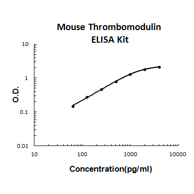
  • For quantitative detection of mouse Thrombomodulin in cell culture supernates, serum, plasma(heparin, EDTA, citrate) and urine.   


    Brand:
    Boster Bio
    SKU:EK1119

    Available on backorder
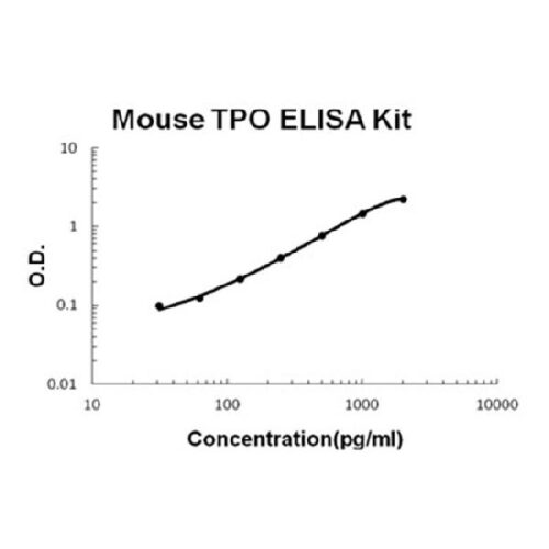
  • For quantitative detection of mouse TPO in cell culture supernates, cell lysates, serum and plasma (heparin, EDTA).   


    Brand:
    Boster Bio
    SKU:EK0517

    Available on backorder
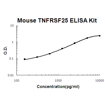
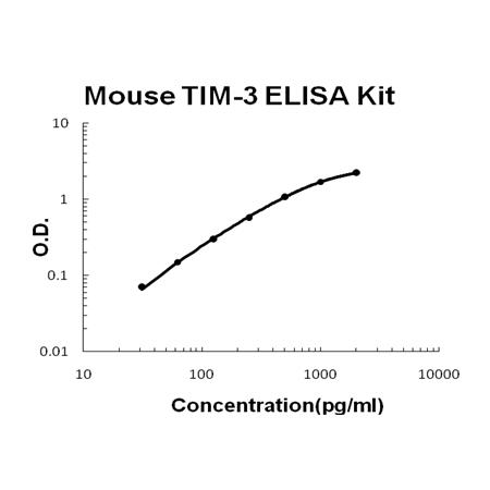
  • For Quantitative Detection of mouse TIM-3 in cell culture supernates, serum and plasma (heparin, EDTA).   


    Brand:
    Boster Bio
    SKU:EK1645

    Available on backorder
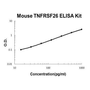
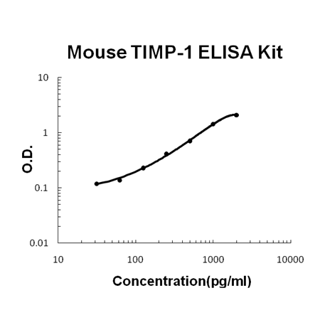
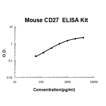
  • For quantitative detection of mouse TIMP-1 in cell culture supernates, cell lysates, serum and plasma (heparin, EDTA).   


    Brand:
    Boster Bio
    SKU:EK0521

    Available on backorder
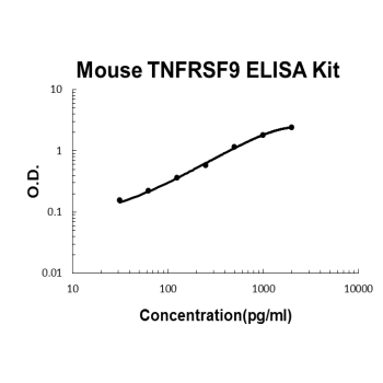
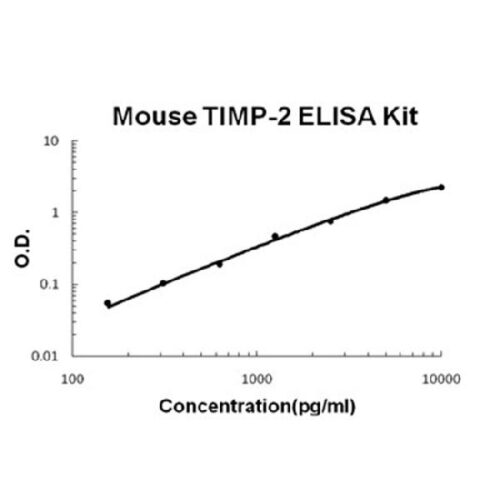
  • For quantitative detection of mouse TIMP-2 in cell culture supernates, cell lysates, serum and plasma (heparin, EDTA).   


    Brand:
    Boster Bio
    SKU:EK0955

    Available on backorder

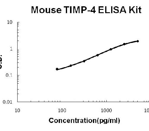
  • For quantitative detection of mouse TIMP-4 in cell culture supernates, cell lysates, serum and plasma (heparin, EDTA).   


    Brand:
    Boster Bio
    SKU:EK1335

    Available on backorder

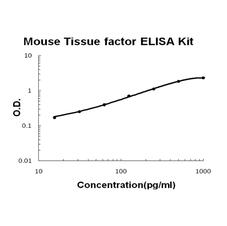
  • For quantitative detection of mouse Tissue factor in cell culture supernates, serum, plasma(heparin, EDTA, citrate) and cell lysates.   


    Brand:
    Boster Bio
    SKU:EK1367

    Available on backorder

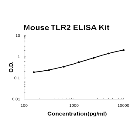
  • For quantitative detection of mouse TLR2 in cell culture supernates, cell lysates, serum and plasma (heparin, EDTA).   


    Brand:
    Boster Bio
    SKU:EK1130

    Available on backorder

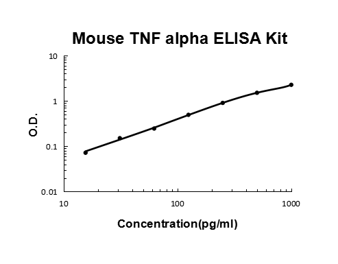
  • For quantitative detection of mouse TNF alpha in cell culture supernates, cell lysates, serum and plasma (heparin, EDTA).   


    Brand:
    Boster Bio
    SKU:EK0527

    Available on backorder

  • For the quantitation of Mouse Tnf concentrations in cell culture supernates, cell lysates, serum and plasma (heparin, EDTA).   


    Brand:
    Boster Bio
    SKU:FEK0527

    Available on backorder

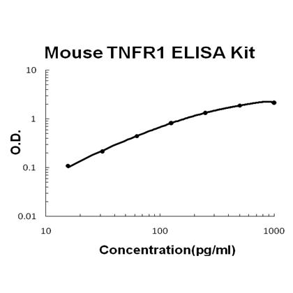
  • For quantitative detection of mouse TNFR1 in cell culture supernates and serum.   


    Brand:
    Boster Bio
    SKU:EK0529

    Available on backorder

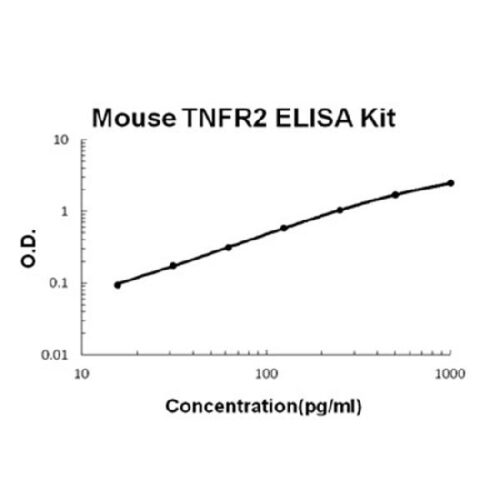
  • For quantitative detection of mouse TNFR2 in cell culture supernates and serum.   


    Brand:
    Boster Bio
    SKU:EK0531

    Available on backorder

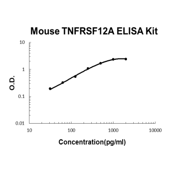
  • For quantitative detection of mouse TNFRSF12A/TWEAKR in cell culture supernates, serum and plasma (heparin, EDTA, citrate).   


    Brand:
    Boster Bio
    SKU:EK1754

    Available on backorder

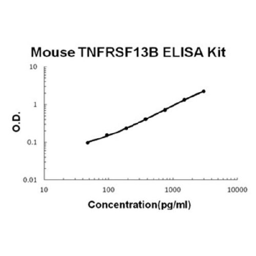
  • For quantitative detection of mouse TNFRSF13B in cell culture supernates, cell lysates, serum and plasma (heparin, EDTA).   


    Brand:
    Boster Bio
    SKU:EK1144

    Available on backorder

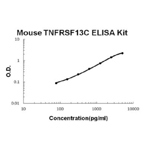
  • For quantitative detection of mouse TNFRSF13C in cell culture supernates, cell lysates, serum and plasma (heparin, EDTA).   


    Brand:
    Boster Bio
    SKU:EK1122

    Available on backorder

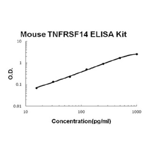
  • For quantitative detection of mouse TNFRSF14 in cell culture supernates, cell lysates, serum and plasma (heparin, EDTA).   


    Brand:
    Boster Bio
    SKU:EK1227

    Available on backorder

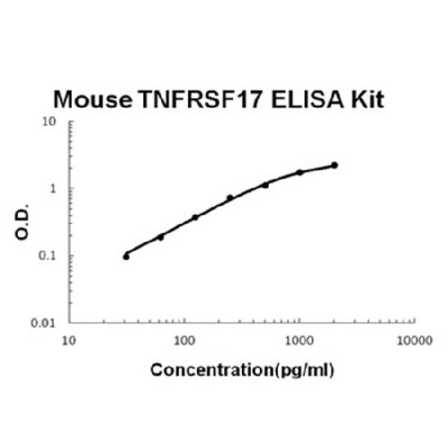
  • For quantitative detection of mouse TNFRSF17 in cell culture supernates, cell lysates, serum and plasma (heparin, EDTA).   


    Brand:
    Boster Bio
    SKU:EK0662

    Available on backorder

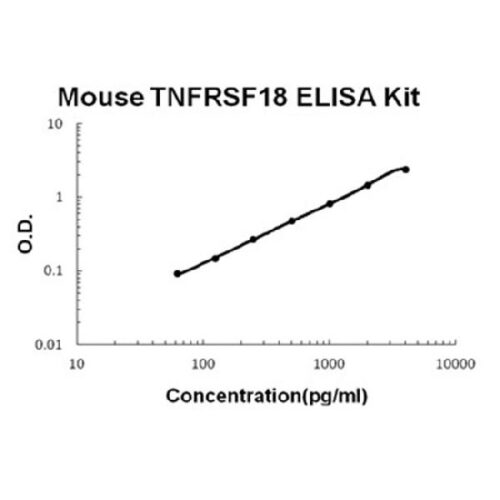
  • For quantitative detection of mouse TNFRSF18 in cell culture supernates, cell lysates, serum and plasma (heparin, EDTA).   


    Brand:
    Boster Bio
    SKU:EK0769

    Available on backorder

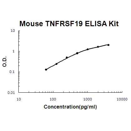
  • For quantitative detection of mouse TNFRSF19 in cell culture supernates, cell lysates, serum and plasma (heparin, EDTA).   


    Brand:
    Boster Bio
    SKU:EK1585

    Available on backorder

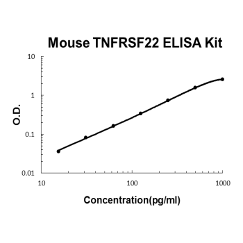
  • For quantitative detection of mouse TNFRSF22 in cell culture supernates, serum and plasma (heparin, EDTA, citrate).   


    Brand:
    Boster Bio
    SKU:EK2007

    Available on backorder

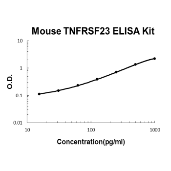
  • For quantitative detection of mouse TNFRSF23 in cell culture supernates, serum and plasma (heparin, EDTA, citrate).   


    Brand:
    Boster Bio
    SKU:EK2008

    Available on backorder

  • For quantitative detection of mouse TNFRSF25 in cell culture supernates, cell lysates, serum and plasma (heparin, EDTA).   


    Brand:
    Boster Bio
    SKU:EK1515

    Available on backorder

  • For quantitative detection of mouse TNFRSF26 in cell culture supernates, serum and plasma (heparin, EDTA, citrate).   


    Brand:
    Boster Bio
    SKU:EK2009

    Available on backorder

  • For quantitative detection of mouse TNFRSF4 in cell culture supernates, cell lysates, serum and plasma (heparin, EDTA)   


    Brand:
    Boster Bio
    SKU:EK0998

    Available on backorder

  • For quantitative detection of mouse TNFRSF7 in cell culture supernates and serum.   


    Brand:
    Boster Bio
    SKU:EK0699

    Available on backorder

  • For quantitative detection of mouse TNFRSF9 in cell culture supernates, cell lysates, serum and plasma (heparin, EDTA).   


    Brand:
    Boster Bio
    SKU:EK1143

    Available on backorder

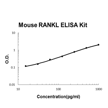
  • For quantitative detection of mouse TNFSF11 in cell culture supernates, cell lysates, serum and plasma (heparin, EDTA).   


    Brand:
    Boster Bio
    SKU:EK0843

    Available on backorder

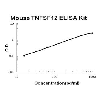
  • For quantitative detection of mouse TNFSF12 in cell culture supernates, cell lysates, serum and plasma (heparin, EDTA).   


    Brand:
    Boster Bio
    SKU:EK1176

    Available on backorder

  • For quantitative detection of mouse TNFSF14 in cell culture supernates, cell lysates, serum and plasma (heparin, EDTA).   


    Brand:
    Boster Bio
    SKU:EK1174

    Available on backorder

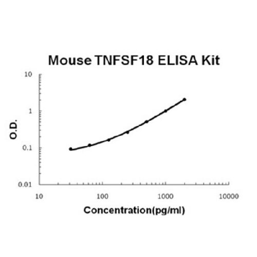
  • For quantitative detection of mouse TNFSF18 in cell culture supernates, cell lysates, serum and plasma (heparin, EDTA).   


    Brand:
    Boster Bio
    SKU:EK1308

    Available on backorder

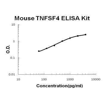
  • For quantitative detection of mouse TNFSF4 in cell culture supernates, cell lysates, serum and plasma (heparin, EDTA).   


    Brand:
    Boster Bio
    SKU:EK0858

    Available on backorder

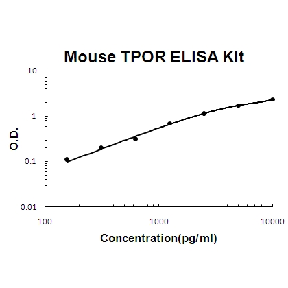
  • For quantitative detection of mouse TPOR in cell culture supernates, cell lysates, serum and plasma (heparin, EDTA).   


    Brand:
    Boster Bio
    SKU:EK1436

    Available on backorder

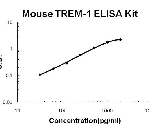
  • For quantitative detection of mouse TREM-1 in cell culture supernates, serum, plasma(heparin, EDTA) and cell lysates.   


    Brand:
    Boster Bio
    SKU:EK0845

    Available on backorder

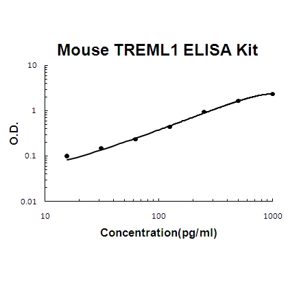
  • For quantitative detection of mouse TREML1 in cell culture supernates, cell lysates, serum and plasma (heparin, EDTA).   


    Brand:
    Boster Bio
    SKU:EK1522

    Available on backorder

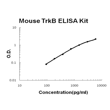
  • For quantitative detection of mouse TrkB in cell culture supernates, cell lysates, serum and plasma (heparin, EDTA).   


    Brand:
    Boster Bio
    SKU:EK0849

    Available on backorder

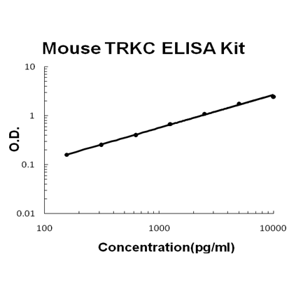
  • For Quantitative Detection of mouse TRKC in cell culture supernates and cell lysates.   


    Brand:
    Boster Bio
    SKU:EK1659

    Available on backorder

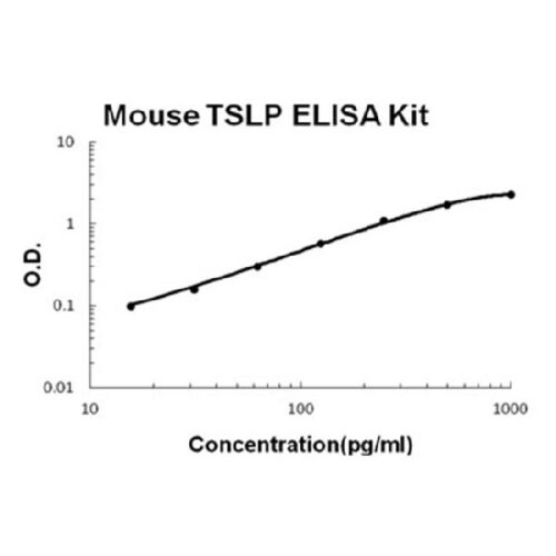
  • For quantitative detection of mouse TSLP in cell culture supernates, cell lysates, serum and plasma (heparin, EDTA).   


    Brand:
    Boster Bio
    SKU:EK1206

    Available on backorder

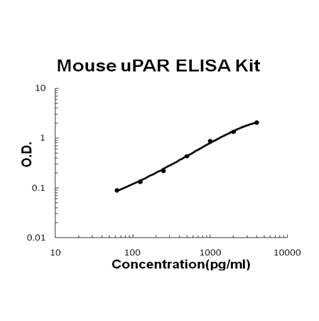
  • For quantitative detection of mouse uPAR in cell culture supernates, cell lysates, serum and plasma (heparin, EDTA).   


    Brand:
    Boster Bio
    SKU:EK1215

    Available on backorder

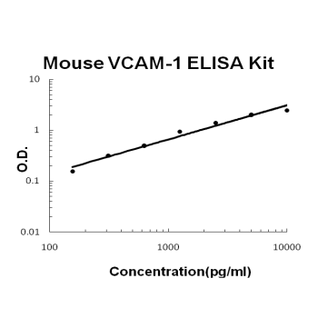
  • For quantitative detection of mouse VCAM-1 in cell culture supernates, cell lysates, serum and plasma (heparin, EDTA).   


    Brand:
    Boster Bio
    SKU:EK0538

    Available on backorder

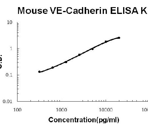
  • For quantitative detection of mouse VE-Cadherin in cell culture supernates, cell lysates, serum and plasma (heparin, EDTA).   


    Brand:
    Boster Bio
    SKU:EK1318

    Available on backorder

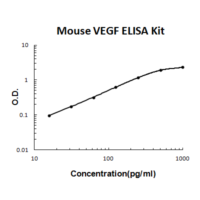
  • For quantitative detection of mouse VEGF in cell culture supernates, serum and plasma(heparin, EDTA, citrate).   


    Brand:
    Boster Bio
    SKU:EK0541

    Available on backorder

  • The Fast version of Picokine ELISA kits, assay takes less than 1.5 hours. Detect Mouse Vegf/VEGFA with <2pg/ml sensitivity. Format: 96-well plate with removable strips. Compatible samples: cell culture supernates, serum and plasma(heparin, EDTA, citrate). This is a TMB colorimetric sandwich ELISA kit with short assay time and fast experiment set up. Vegf/VEGFA tissue specificity: In developing embryos, expressed mainly in the choroid plexus, paraventricular neuroepithelium, placenta and kidney glomeruli. Also found in bronchial epithelium, adrenal gland and in seminiferous tubules of testis. High expression of VEGF continues in kidney glomeruli and choroid plexus in adults.   


    Brand:
    Boster Bio
    SKU:FEK0541

    Available on backorder

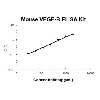
  • For quantitative detection of mouse VEGF-B in cell culture supernates, serum and plasma(heparin).   


    Brand:
    Boster Bio
    SKU:EK1411

    Available on backorder

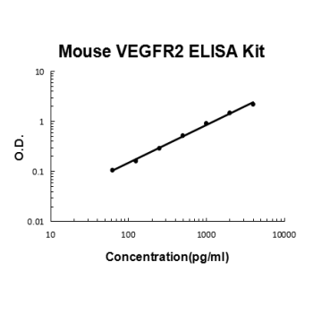
  • For quantitative detection of mouse VEGFR2 in cell culture supernates and serum.   


    Brand:
    Boster Bio
    SKU:EK0590

    Available on backorder

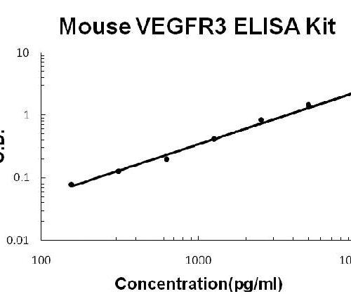
  • For quantitative detection of mouse VEGFR3 in cell culture supernates and serum.   


    Brand:
    Boster Bio
    SKU:EK0546

    Available on backorder

  • For quantitative detection of mouse Vitamin DBP in cell culture supernates, lysates, serum, plasma (heparin) and urine.   


    Brand:
    Boster Bio
    SKU:EK2063

    Available on backorder

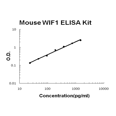
  • For quantitative detection of mouse WIF1 in cell culture supernates, cell lysates, serum and plasma (heparin, EDTA).   


    Brand:
    Boster Bio
    SKU:EK1523

    Available on backorder

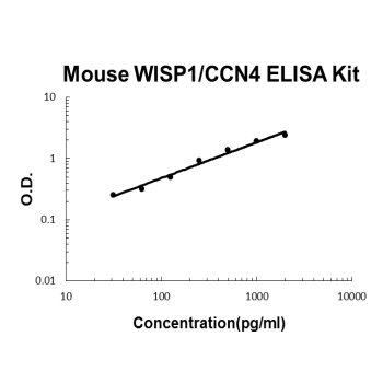
  • For quantitative detection of mouse WISP1 in cell culture supernates, cell lysates, serum and plasma (heparin, EDTA).   


    Brand:
    Boster Bio
    SKU:EK1195

    Available on backorder

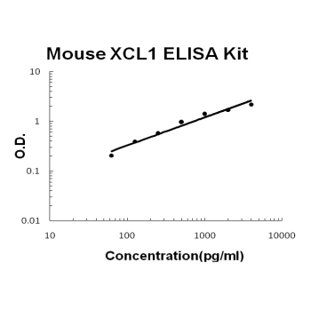
  • For quantitative detection of mouse XCL1 in cell culture supernates, cell lysates, serum and plasma (heparin, EDTA).   


    Brand:
    Boster Bio
    SKU:EK0803

    Available on backorder

  • Brand:
    Cayman
    SKU:700518 - 2 g

    Available on backorder

  • Brand:
    Cayman
    SKU:700164 - 1 ea

    Available on backorder

  • Cayman’s MTT Proliferation Assay Kit provides an easy to use tool for studying the induction and inhibition of cell proliferation in any in vitro model. This kit will also allow investigators to screen drug candidates involved in cell cycle regulation. In this assay, MTT is taken up by cells through the plasma membrane potential and then reduced to formazan by intracellular NAD(P)H-oxidoreductases.{14422}  


    Brand:
    Cayman
    SKU:10009365 - 480 wells

    Available on backorder

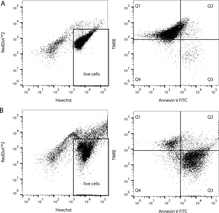
  • Cayman’s Multi-Parameter Apoptosis Assay Kit is a simple way to probe for multiple different apoptotic readouts at the same time. This kit employs FITC-conjugated Annexin V as a probe for phosphatidylserine on the outer membrane of apoptotic cells, TMRE as a probe for Δψm, RedDotTM2 as an indicator of plasma membrane permeability/cell viability, and Hoechst Dye to demonstrate nuclear morphology. The kit allows phenotypic characterization of multiple different cell death parameters at the single-cell level. The reagents provided in the kit are sufficient to run 100 samples.  


    Brand:
    Cayman
    SKU:601280 - 100 tests

    Available on backorder

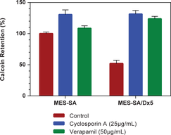
  • Cayman’s Multidrug Resistance Assay Kit provides a convenient tool for studying the modulation of cellular multidrug resistance (MDR) machinery. The kit employs calcein AM, a cell-permeable nonfluorescent dye, which is cleaved intracellularly to yield the impermeant fluorescent molecule, calcein. This calcein is rapidly excluded from cells expressing the MDR proteins P-gp or MRP, and so functions as a probe for the detection of chemical compounds inhibiting MDR proteins. Propidium iodide is provided as a stain for marking dead or damaged cells. Cyclosporin A, a competitive inhibitor of MDR proteins, and verapamil, a noncompetitive inhibitor, are included as controls.  


    Brand:
    Cayman
    SKU:600370 - 1,000 tests

    Available on backorder

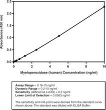
  • Myeloperoxidase (MPO) is a peroxidase enzyme with roles in innate immunoresponse and various diseases. Cayman’s MPO (human) ELISA Kit is a sandwich assay that can be used to measure MPO in plasma and serum without prior sample purification. This assay has been validated using plasma and serum from healthy volunteers and has also been validated in RPMI-1640 with 10% fetal bovine serum. The standard curve spans the range of 0.16-10 ng/ml and has a sensitivity (defined as LLOQ) of 0.2 ng/ml.  


    Brand:
    Cayman
    SKU:501410 - 96 wells

    Available on backorder

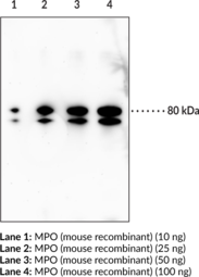
  • Myeloperoxidase (MPO) is a heme-containing enzyme and the most abundant protein in polymorphonuclear leukocytes (PMNs).{57128} It is comprised of two subunits linked by a disulfide bridge with each subunit containing a light and a heavy polypeptide chain. It can oxidize a variety of substrates and catalyzes the formation of highly reactive (pseudo)hypohalous acids and radicals including hypochlorous acid. MPO is stored in azurophilic granules of PMNs and is released from activated or necrotic PMNs, after which it can bind to and modify acidic serum proteins, as well as recruit additional PMNs. MPO also has roles in PMN apoptosis and antimicrobial defense systems, including neutrophil extracellular traps (NETs).{57128,23608,24590} MPO-deficient mice exhibit reduced survival in a polymicrobial sepsis model, increased susceptibility to experimental autoimmune encephalomyelitis (EAE), and increased atherosclerosis in mice also deficient in the LDL receptor and fed an atherogenic diet.{57128,57129,57127} Cayman’s Myeloperoxidase (mouse) Polyclonal Antibody can be used for ELISA, immunohistochemistry (IHC), and Western blot (WB) applications. The antibody recognizes MPO at approximately 80 kDa from mouse samples.  


    Brand:
    Cayman
    SKU:20493 - 1 ea

    Available on backorder

  • Brand:
    Cayman
    SKU:700166 - 1 ea

    Available on backorder

  • Cayman’s Myeloperoxidase Chlorination Fluorometric Assay provides a convenient fluorescence-based method for detecting the MPO chlorination activity in both crude cell lysates and purified enzyme preparations. The assay utilizes the non-fluorescent 2-[6-(4-aminophenoxy)-3-oxo-3H-xanthen-9-yl]-benzoic acid (APF), which is selectively cleaved by hypochlorite (-OCl) to yield the highly fluorescent compound fluorescein.{11242} Fluorescein fluorescence is analyzed with an excitation wavelength of 480-495 nm and an emission wavelength of 515-525 nm. The kit includes a MPO-specific inhibitor for distinguishing MPO activity from MPO-independent fluorescence.  


    Brand:
    Cayman
    SKU:10006438 - 2 x 96 wells

    Available on backorder

  • Myeloperoxidase (MPO) is a member of the heme peroxidase superfamily and is stored within the azurophilic granules of leukocytes.{16902} MPO is found within circulating neutrophils, monocytes, and some tissue macrophages.{16923} A unique activity of MPO is its ability to use chloride as a cosubstrate with hydrogen peroxide to generate chlorinating oxidants such as hypochlorous acid, a potent antimicrobial agent.{16904} Recently, evidence has emerged that MPO-derived oxidants contribute to tissue damage and the initiation and propagation of acute and chronic vascular inflammatory diseases.{9886,9889} The fact that circulating levels of MPO have been shown to predict risks for major adverse cardiac events and that levels of MPO-derived chlorinated compounds are specific biomarkers for disease progression, has attracted considerable interest in the development of therapeutically useful MPO inhibitors.{16905} MPO also oxidizes a variety of substrates, including phenols and anilines, via the classic peroxidation cycle. The relative concentrations of chloride and the reducing substrate determine whether MPO uses hydrogen peroxide for chlorination or peroxidation. Assays based on measurement of chlorination activity are more specific for MPO than those based on peroxidase substrates because peroxidases generally do not produce hypochlorous acid. However, it is important that when screening for MPO inhibition that both the chlorination and peroxidation activities be tested. This determines whether the inhibitor specifically interferes with the chlorination and/or peroxidation cycle or whether the inhibitor simply acts as a scavenger for hypochlorous acid. Also, many reversible inhibitors act by diverting MPO from the chlorinating cycle to the peroxidase cycle. Cayman’s MPO Inhibitor Screening Assay provides convenient fluorescence-based methods for screening inhibitors to both the chlorination and peroxidation activities of MPO. The chlorination assay utilizes the non-fluorescent 2-[6-(4-aminophenoxy)-3-oxo-3H-xanthen-9-yl]-benzoic acid (APF), which is selectively cleaved by hypochlorite (-OCl) to yield the highly fluorescent compound fluorescein.{11242} Fluorescein fluorescence is analyzed with an excitation wavelength of 480-495 nm and an emission wavelength of 515-525 nm. The peroxidation assay utilizes the peroxidase component of MPO. The reaction between hydrogen peroxide and ADHP (10-acetyl-3,7-dihydroxyphenoxazine) produces the highly fluorescent compound resorufin. Resorufin fluorescence is analyzed with an excitation wavelength of 530-540 nm and an emission wavelength of 585-595 nm.  


    Brand:
    Cayman
    SKU:700170 - 2 x 96 wells

    Available on backorder

  • Myeloperoxidase (MPO) is a heme-containing enzyme and the most abundant protein in polymorphonuclear leukocytes (PMNs).{57128} It is comprised of two subunits linked by a disulfide bridge with each subunit containing a light and a heavy polypeptide chain. It can oxidize a variety of substrates and catalyzes the formation of highly reactive (pseudo)hypohalous acids and radicals, including hypochlorous acid. MPO is stored in azurophilic granules of PMNs and is released from activated or necrotic PMNs, after which it can bind to and modify acidic serum proteins, as well as recruit additional PMNs. MPO also has roles in PMN apoptosis and antimicrobial defense systems, including neutrophil extracellular traps (NETs).{57128,23608,24590} MPO-deficient mice exhibit reduced survival in a polymicrobial sepsis model, increased susceptibility to experimental autoimmune encephalomyelitis (EAE), and increased atherosclerosis in mice also deficient in the LDL receptor and fed an atherogenic diet.{57128,57129,57127} Cayman’s Myeloperoxidase Monoclonal Antibody (Clone 2C8) was developed by fusing the spleen of a non-immunized NZBWF1 mouse with a mouse myeloma cell line and can be used for immunofluorescence (IF) applications.  


    Brand:
    Cayman
    SKU:15638 - 1 ea

    Available on backorder

  • Myeloperoxidase (MPO) is a heme-containing enzyme and the most abundant protein in polymorphonuclear leukocytes (PMNs).{57128} It is comprised of two subunits linked by a disulfide bridge with each subunit containing a light and a heavy polypeptide chain. It can oxidize a variety of substrates and catalyzes the formation of highly reactive (pseudo)hypohalous acids and radicals, including hypochlorous acid. MPO is stored in azurophilic granules of PMNs and is released from activated or necrotic PMNs, after which it can bind to and modify acidic serum proteins, as well as recruit additional PMNs. MPO also has roles in PMN apoptosis and antimicrobial defense systems, including neutrophil extracellular traps (NETs).{57128,23608,24590} MPO-deficient mice exhibit reduced survival in a polymicrobial sepsis model, increased susceptibility to experimental autoimmune encephalomyelitis (EAE), and increased atherosclerosis in mice also deficient in the LDL receptor and fed an atherogenic diet.{57128,57129,57127} Cayman’s Myeloperoxidase Monoclonal Antibody (Clone 5F6) was developed by fusing the spleen of a non-immunized NZBWF1 mouse with a mouse myeloma cell line and can be used for immunofluorescence (IF) applications.  


    Brand:
    Cayman
    SKU:15640 - 1 ea

    Available on backorder

  • Myeloperoxidase (MPO) is a member of the heme peroxidase superfamily and is stored within the azurophilic granules of leukocytes.{16902} MPO is found within circulating neutrophils, monocytes, and some tissue macrophages.{16923} A unique activity of MPO is its ability to use chloride as a cosubstrate with hydrogen peroxide to generate chlorinating oxidants such as hypochlorous acid, a potent antimicrobial agent.{16904} Recently, evidence has emerged that MPO-derived oxidants contribute to tissue damage and the initiation and propagation of acute and chronic vascular inflammatory diseases.{9886,9889} The fact that circulating levels of MPO have been shown to predict risks for major adverse cardiac events and that levels of MPO-derived chlorinated compounds are specific biomarkers for disease progression, has attracted considerable interest in the development of therapeutically useful MPO inhibitors.{16905} MPO also oxidizes a variety of substrates, including phenols and anilines, via the classic peroxidation cycle. The relative concentrations of chloride and the reducing substrate determine whether MPO uses hydrogen peroxide for chlorination or peroxidation. Cayman’s MPO Peroxidation Fluorometric Assay provides a convenient fluorescence-based method for detecting the MPO peroxidase activity in both crude cell lysates and purified enzyme preparations. The assay utilizes the peroxidase component of MPO. The reaction between hydrogen peroxide and ADHP (10-acetyl-3,7-dihydroxyphenoxazine) produces the highly fluorescent compound resorufin. Resorufin fluorescence can be easily analyzed with an excitation wavelength of 530-540 nm and emission wavelength of 585-595 nm. The kit includes a MPO-specific inhibitor for distinguishing between MPO activity from MPO-independent fluorescence.  


    Brand:
    Cayman
    SKU:700160 - 2 x 96 wells

    Available on backorder

  • N6-Methyladenosine (m6A) is an abundant modification found in mRNA, tRNA, snRNA, as well as long non-coding RNA, in all species. RNA adenosine methylation is catalyzed by a multicomponent complex composed of METTL3/MT-A70, METTL14, and WTAP in mammals. METTL3 and METTL14 are responsible for the methyltransferase activity of the complex, and WTAP mediates substrate recruitment.{30057} The process shares similarities to histone methylation in that the modification is installed by m6A methylation “writers”, detected by methylation-specific “readers”, and can be reversed by demethylation “erasers”. The m6A modification has been shown to be conserved in the vicinity of stop codons and the 3’-untranslated region of specific mouse and human mRNAs.{25677} The process of regulating m6A modifications in mammalian mRNA has been linked to disease, where fat mass and obesity-associated (FTO) has been reported to be an obesity risk gene.{25677} FTO is an m6A demethylase and polymorphisms that result in increased FTO expression are associated with increased body mass and risk of obesity. Cayman’s N6-Methyladenosine Monoclonal Antibody (Clone 17-3-4-1) can be used for dot blot, ELISA, and immunoprecipitation applications.  


    Brand:
    Cayman
    SKU:18336 - 100 µg

    Available on backorder

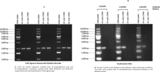
  • N6-Methyladenosine (m6A) is the most prevalent internal modification that occurs in the messenger RNAs (mRNAs) of most eukaryotes and has been linked to effects on mRNA fate. The process shares similarities to histone methylation in that the modification is installed by m6A methylation “writers”, detected by methylation-specific “readers”, and can be reversed by demethylation “erasers”. The m6A modification has been found to be highly conserved around stop codons, in 3’-untranslated region, and within long external exons in both human and mouse cells.{25677} The process of regulating m6A modifications in mammalian mRNA has been linked to disease, where fat mass and obesity-associated (FTO) has been reported to be an obesity risk gene.{25677} FTO is an m6A demethylase and polymorphisms that result in increased FTO expression are associated with increased body mass and risk of obesity. Cayman’s N6-Methyladenosine Polyclonal Antibody can be used for ELISA and Southwestern dot blot applications.  


    Brand:
    Cayman
    SKU:18337 - 1 ea

    Available on backorder

  • Nicotinamide adenine dinucleotide (NAD) exists in an oxidized form, NAD+, as well as a reduced form, NADH. NAD, the main free form in cells, functions in modulating cellular redox status and by controlling signaling and transcriptional events, making it an important cofactor when investigating normal cellular function. Cayman’s NAD/NADH Cell-Based Assay Kit provides a colorimetric method for measuring intracellular NAD+ and NADH in cultured cells. In this assay, NAD+ found in cell samples is reduced to NADH by alcohol dehydrogenase during the oxidation of ethanol to acetaldehyde. The newly formed and the existing NADH found in the samples is then oxidized resulting in the reduction of a tetrazolium salt substrate (WST-1) to a highly-colored formazan which absorbs at 450 nm. The amount of formazan produced is proportional to the amount of total NAD in the cell lysate and can be used as an indicator of the total cellular NAD concentration.  


    Brand:
    Cayman
    SKU:600480 - 1 ea

    Available on backorder

  • Brand:
    Cayman
    SKU:700932 - 500 µg

    Available on backorder

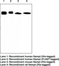
  • Nampt is a 52 kDa adipokine secreted by adipose tissue that is involved in the biosynthesis of nicotinamide adenine dinucleotide (NAD+). Two forms of Nampt exist, an intracellular form (iNampt) and an extracellular form (eNampt). While the function of iNampt as an essential and rate-limiting NAD+ biosynthetic enzyme is well established, the physiological role of eNampt is still a matter of debate. Nampt has various functions, including the promotion of vascular smooth muscle cell maturation and inhibition of neutrophil apoptosis. It activates the insulin receptor and has insulin-mimetic effects, lowering blood glucose and improving insulin sensitivity. The protein is highly expressed in visceral fat and serum levels of the protein correlate with obesity.  


    Brand:
    Cayman
    SKU:10813 - 100 µg

    Available on backorder

  • Nampt is a 52 kDa adipokine secreted by adipose tissue that is involved in the biosynthesis of nicotinamide adenine dinucleotide (NAD+). Two forms of Nampt exist, an intracellular form (iNampt) and an extracellular form (eNampt). While the function of iNampt as an essential and rate-limiting NAD+ biosynthetic enzyme is well established, the physiological role of eNampt is still a matter of debate. Nampt has various functions, including the promotion of vascular smooth muscle cell maturation and inhibition of neutrophil apoptosis. It activates the insulin receptor and has insulin-mimetic effects, lowering blood glucose and improving insulin sensitivity. The protein is highly expressed in visceral fat and serum levels of the protein correlate with obesity.  


    Brand:
    Cayman
    SKU:10813 - 50 µg

    Available on backorder

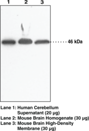
  • N-acylethanolamines (NAEs) are involved in diverse biological processes such as inflammatory regulation, apoptosis, and tissue degeneration.{12164} In animals, NAEs are mainly biosynthesized via a membrane phospholipid-dependent pathway, which is the enzymatic hydrolysis of N-acyl-phosphatidylethanolamine (NAPE). The enzyme catalyzing this reaction is a phospholipase D subtype selective for NAPE named N-acyl-phosphatidylethanolamine-hydrolysing phospholipase D (NAPE-PLD). It has been cloned from mouse, rat, and human and is 393-396 amino acids in length, with an estimated molecular weight of 46 kDa. Both NAPE-PLD mRNA and protein activity have been detected in a wide range of tissues with the highest levels in brain, kidney, and testis.{11963} In rat, NAPE-PLD activity in the brain is low in neonates and is 15-fold higher in adults, whereas the activity remains constant in the heart during development.{13170}  


    Brand:
    Cayman
    SKU:10305 - 1 ea

    Available on backorder

  • N-acylethanolamines (NAEs) are involved in diverse biological processes such as inflammatory regulation, apoptosis, and tissue degeneration.{12164} In animals, NAEs are mainly biosynthesized via a membrane phospholipid-dependent pathway, which is the enzymatic hydrolysis of N-acyl-phosphatidylethanolamine (NAPE). The enzyme catalyzing this reaction is a phospholipase D subtype selective for NAPE named N-acyl-phosphatidylethanolamine-hydrolysing phospholipase D (NAPE-PLD). It has been cloned from mouse, rat, and human and is 393-396 amino acids in length, with an estimated molecular weight of 46 kDa. Both NAPE-PLD mRNA and protein activity have been detected in a wide range of tissues with the highest levels in brain, kidney, and testis.{11963} In rat, NAPE-PLD activity in the brain is low in neonates and is 15-fold higher in adults, whereas the activity remains constant in the heart during development.{13170}  


    Brand:
    Cayman
    SKU:10306 - 500 µl

    Available on backorder

  • Nav1.7 is a voltage-gated sodium channel encoded by the SCN9A gene that is expressed in sensory and dorsal root ganglia neurons as well as in the superficial laminae of the spinal cord.{41708} Nav1.7 amplifies small depolarizations of the membrane and is involved in pain perception. Inactivating gene mutations for Nav1.7 are responsible for congenital insensitivity to pain, a disorder characterized by a complete lack of pain perception. Activating gene mutations for Nav1.7 are responsible for various pain-related disorders. Mutations that decrease the action potential threshold by shifting the voltage dependence and slowing deactivation lead to hyperexcitability of dorsal root ganglia neurons in inherited erythromelalgia, which is characterized by redness and burning sensations in the feet.{41709} Missense mutations that reduce fast inactivation of Nav1.7 channels lead to persistent sodium current and are associated with paroxysmal pain disorder, which affects the sacral region and face.{41710} Cayman’s Nav1.7 Monoclonal Antibody (Clone S68-6) can be used for Western blot, immunoprecipitation, and immunocytochemistry applications.  


    Brand:
    Cayman
    SKU:13718 - 100 µg

    Available on backorder

  • CD56, also known as neural cell adhesion molecule 1 (NCAM1), is a cell surface glycoprotein and member of the immunoglobulin superfamily that is involved in cellular adhesion and neural development.{60124,60125,60126} Alternative splicing of the NCAM1 pre-mRNA produces several isoforms, including two transmembrane isoforms of 180 and 140 kDa, NCAM-180 and NCAM-140, respectively, that differ in size of the cytoplasmic domain, and a 120 kDa isoform, NCAM-120, that lacks the cytoplasmic domain and is linked to the plasma membrane by a glycosylphosphatidylinositol (GPI) anchor.{60124} CD56 is composed of an extracellular N-terminal domain containing five immunoglobulin-like (Ig-like) domains and two fibronectin type III domains that mediate homophilic and heterophilic interactions, a transmembrane domain, and an intracellular C-terminal signaling domain.{60125,60127} It is used as a marker for neural and natural killer (NK) cells, but is also expressed by other immune cells, including monocytes, T cells, and dendritic cells (DCs), as well as tumor cells, in an isoform-dependent manner.{60124,60125,60127} CD56 is increased on NK cells by stimulation with IL-15 or IL-2 and correlates with upregulation of the NK cell activating receptors NKG2D, NKp30, and NKp46 and NK cell-mediated cytotoxicity.{60124,60127} Homophilic CD56 interactions induce neurite outgrowth of cerebellar neurons and promote NK cell-mediated tumor cell lysis in vitro.{60124,60125,60126,60127} The frequency of peripheral blood CD56bright NK cells is increased in patients with melanoma and associated with reduced overall survival.{60128} Cayman’s NCAM1/CD56 (C-Term) Rabbit Monoclonal Antibody can be used for immunohistochemistry (IHC) and Western blot (WB) applications. The antibody recognizes NCAM-180 and NCAM-140 from human samples.  


    Brand:
    Cayman
    SKU:32257 - 100 µl

    Available on backorder

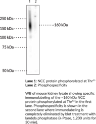
  • The thiazide-sensitive sodium chloride cotransporter (NCC) is a member of the SLC12 family of transporters and is encoded by SLC12A3 in humans.{46961} NCC is expressed in epithelial cells of the distal convoluted tubule and localizes to the apical plasma membrane where it mediates sodium reabsorption in the kidney. It consists of a central hydrophobic domain with 12 transmembrane helices that contain affinity-modifying residues for sodium, chloride, and thiazides that is flanked by intracellular N- and C-terminal domains with sites that are subject to phosphorylation. Phosphorylation of NCC at threonine 53 (Thr53) is mediated by serum- and glucocorticoid-inducible kinase 1 (SGK1), STE20/SPS1-related proline-alanine-rich protein kinase (SPAK), or oxidative stress-responsive kinase 1 (OSR1). X. laevis oocytes expressing a threonine-to-alanine substitution at Thr53 in NCC, which abolishes its phosphorylation, have reduced chloride deprivation-induced sodium uptake, but not NCC surface expression, compared to wild-type oocytes.{46963} NCC (phospho-Thr53) levels are increased in the kidney by low dietary intake of sodium chloride or potassium in mice.{46962} NCC (phospho-Thr53) levels are also increased in the kidney of the CUL3-Het/∆9 mouse model of familial hyperkalemic hypertension.{46964} Cayman’s NCC (Phospho-Thr53) Polyclonal Antibody can be used for immunofluorescence (IF) and Western blot (WB) applications. The antibody recognizes NCC (phospho-Thr53) at approximately 160 kDa from human, mouse, and rat samples.  


    Brand:
    Cayman
    SKU:29285 - 100 µl

    Available on backorder

  • [Bertin Catalog No. G01021]  


    Brand:
    Cayman
    SKU:32790 - 100 µl

    Available on backorder

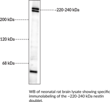
  • Nestin, also known as neuroepithelial stem cell protein, is a type VI intermediate filament protein.{55198} Nestin monomers are comprised of a short N-terminal head domain that is required for assembly of intermediate filaments, an α-helical rod domain that mediates dimerization, and a long C-terminal tail.{55198} They form heterodimers and heterotetramers with other intermediate filament proteins, including vimentin and α-internexin, for assembly into intermediate filaments.{55198,55199} Nestin is found in a variety of tissue types but is expressed primarily in nervous tissue during embryonic development in mammals and has been used as a marker of neural stem/progenitor cells (NSPCs).{55199,55200} Nestin synthesis decreases as nervous tissues differentiate, however, nestin-positive neural stem cells are found in the subventricular zone (SVZ) and hippocampal dentate gyrus in the adult mammalian brain. Nestin is also expressed in a variety of tumor types and cancer stem cells, as well as in cerebellar Purkinje cells from patients with Creutzfeldt-Jakob disease (CJD).{55200,55201} Cayman’s Nestin Monoclonal Antibody can be used for immunocytochemistry (ICC), immunohistochemistry (IHC), and Western blot (WB) applications. The antibody recognizes a nestin doublet at approximately 220 to 240 kDa from human, mouse, and rat samples.  


    Brand:
    Cayman
    SKU:29286 - 100 µl

    Available on backorder

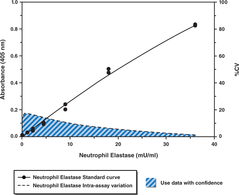
  • Cayman’s NETosis Assay Kit provides a simple and fast method for studying the process of NETosis ex vivo. Notably, Cayman’s NETosis Assay Kit does not depend upon the DNA component of neutrophil extracellular traps (NETs), as DNA release can occur independently of NETosis. In this kit, primary neutrophils are stimulated to release NETs with either PMA or a calcium ionophore (both included). Unbound neutrophil elastase is washed away following NET generation. Following digest of NET DNA by S7 nuclease, the supernatant containing neutrophil elastase is added to a substrate, which is selectively cleaved by elastase to yield a 4-nitroaniline product that adsorbs light at 405 nm. Enough reagents are provided to test 24 sample conditions for NET production, with analysis in duplicate.  


    Brand:
    Cayman
    SKU:601010 - 1 ea

    Available on backorder

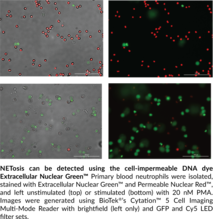
  • Cayman’s NETosis Imaging Kit provides a simple staining protocol for visualizing the process of NETosis kinetically ex vivo and in vitro. A cell-permeable DNA dye allows visualization of the dynamic changes of the nucleus during the process of NETosis, while a cell-impermeable dye detects the extruded DNA. Combining the fluorescence visualization of these dyes with brightfield imaging in a high-content platform allows for exquisite differentiation between different modes of cell death. The amount of each reagent provided is sufficient for two 96-well plates.  


    Brand:
    Cayman
    SKU:601750 - 96 wells

    Available on backorder

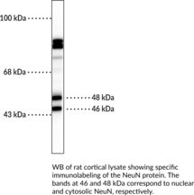
  • NeuN, also known as FOX3 and RNA binding protein fox-1 homolog 3 (Rbfox3), is a pre-mRNA alternative splicing regulator encoded by RBFOX3 in humans.{53699,53701} It is expressed in mature neurons of the brain and spinal cord and is commonly used as a neuronal marker to quantify the number of new neurons generated during adult neurogenesis or the extent of therapeutic neuroprotection in animal models of neurodegenerative disease.{53702,53700} There are two subtypes of NeuN, a 46 kDa nuclear form and 48 kDa cytoplasmic form. Cytosolic NeuN is increased in the lumbar spinal cord in a mouse model of amyotrophic lateral sclerosis (ALS) compared with control animals.{53701} Exon deletions and truncations of NeuN are found in patients with Rolandic epilepsy and RBFOX3 is located within the apparently balanced chromosomal rearrangement (ABCR) regions of chromosomes in patients with developmental delays and speech disorders.{53700} Cayman’s NeuN Monoclonal Antibody can be used for immunocytochemistry (ICC), immunohistochemistry (IHC), and Western blot (WB) applications. The antibody recognizes nuclear and cytosolic NeuN at approximately 46 and 48 kDa, respectively.  


    Brand:
    Cayman
    SKU:29265 - 100 µl

    Available on backorder

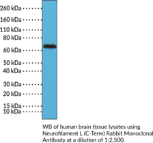
  • Neurofilament L (NF-L) is one of four subunits that form NFs, which are type III intermediate filament proteins that enable axonal growth, maintain structure, and facilitate nerve conduction.{59461,59462} NF-L is comprised of an N-terminal domain that regulates neurofilament assembly, a central α-helical rod region that mediates dimerization, and a glutamic acid-rich C-terminal tail that is subject to phosphorylation.{59461,59463,59462} It is abundantly expressed in myelinated axons in the central and peripheral nervous systems and localizes to the cytoplasm, where it associates with an NF-middle (NF-M) or -heavy (NF-H) subunit to form parallel coiled-coil heterodimers.{59463} These heterodimers then associate in an anti-parallel manner to form tetramers, which assemble into NFs. Cerebrospinal fluid (CSF) NF-L levels have been used as a marker of axonal injury in a variety of neurodegenerative diseases, including amyotrophic lateral sclerosis (ALS), multiple sclerosis (MS), Parkinson’s disease, and Alzheimer’s disease.{59461} Mutations in NEFL, the gene encoding NF-L, have been found in patients with Charcot-Marie-Tooth disease type 2E (CMT2E), a neurological disease characterized by muscle weakness and atrophy.{59464} Cayman’s Neurofilament L (C-Term) Rabbit Monoclonal Antibody can be used for immunohistochemistry (IHC) and Western blot (WB) applications.  


    Brand:
    Cayman
    SKU:32230 - 100 µl

    Available on backorder

  • Neutrophil elastase is stored within cytoplasmic azurophilic granules in the neutrophil and released upon stimulation by pathogens where it acts either as free protein or is associated with networks of extracellular traps (NET). Together with other proteases released from activated neutrophils, neutrophil elastase plays a critical role in degrading invading pathogens and thus provides the earliest line of defense in the immune system. It is thought to play a critical role in tumor invasion and metastasis as well as other inflammatory conditions. Cayman’s Neutrophil Elastase Activity Assay kit is designed to be used to study compounds regulating elastase release in neutrophils. The kit employs a specific non-fluorescent elastase substrate, (Z-Ala-Ala-Ala-Ala)2Rh110, which is selectively cleaved by elastase to yield the highly fluorescent compound R110, which can be analyzed with an excitation wavelength of 485 nm and emission wavelength of 525 nm. Reagents needed to isolate neutrophils from whole blood are included in the kit, as is PMA, which is known to stimulate elastase release from neutrophils.  


    Brand:
    Cayman
    SKU:600610 - 2 x 96 wells

    Available on backorder

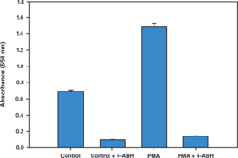
  • Myeloperoxidase (MPO), released from neutrophils to degrade invading pathogens, provides one of the earliest lines of defense in innate immunity. It catalyzes the hydrogen peroxide-mediated oxidation of halide ions to products such as hypochlorous acid (HOCl). These reactive products participate in a variety of secondary reactions which in turn lead to modifications of proteins and destruction of the extracellular matrix. Cayman’s Neutrophil Myeloperoxidase Activity Assay kit utilizes 3,3′,5,5′-tetramethyl-benzidine (TMB) as a chromogenic substrate, which upon reacting with MPO, yields a blue color detectable by its absorbance at 650 nm. The color intensity is proportional to the amount of MPO in the sample. Reagents needed to isolate neutrophils from whole blood are included in the kit, as is PMA, which is known to stimulate MPO release from neutrophils. A specific inhibitor for MPO, 4-aminobenzhydrazide (ABH), is also included in the kit for verifying the specificity of the assay.  


    Brand:
    Cayman
    SKU:600620 - 2 x 96 wells

    Available on backorder

  • Respiratory burst, or the rapid generation of reactive oxygen species from immune cells, is crucial for the destruction of invading microorganisms by phagocytes. During this process, professional phagocytes convert molecular oxygen to superoxide anions through the action of NADPH oxidase, which is then converted to reactive oxygen species with potent antimicrobial activity. Cayman’s Neutrophil/Monocyte Respiratory Burst Assay Kit provides PMA, dihydrorhodamine 123, and additional reagents necessary for inducing and quantifying a respiratory burst response in neutrophils and monocytes by flow cytometry. The assay can be performed on whole blood or on cells in various types of cell culture media. Because the assay reagents are not species-specific, this assay can be used in any species or cell type capable of producing a NADPH oxidase-dependent respiratory burst response.  


    Brand:
    Cayman
    SKU:601130 - 1 ea

    Available on backorder

  • Cayman’s NF-κB (human p50) Transcription Factor Assay is a non-radioactive, sensitive method for detecting specific transcription factor DNA binding activity in nuclear extracts and whole cell lysates. A 96-well enzyme-linked immunosorbent assay (ELISA) replaces the cumbersome radioactive electrophoretic mobility shift assay (EMSA). A specific double stranded DNA (dsDNA) sequence containing the NF-κB response element is immobilized to the wells of a 96-well plate. NF-κB contained in a nuclear extract, binds specifically to the NF-κB response element. NF-κB (p50) is detected by addition of specific primary antibody directed against NF-κB (p50). A secondary antibody conjugated to HRP is added to provide a sensitive colorimetric readout at 450 nm. The Cayman Chemical NF-κB (human p50) Transcription Factor Assay detects human NF-κB (p50). It will not cross-react with NF-κB (p65).  


    Brand:
    Cayman
    SKU:10006912 - 96 wells

    Available on backorder

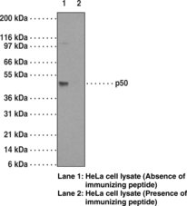
  • NF-κB is a transcription factor activated by various extra and intracellular stimuli such as cytokines, UV radiation, stress, injury, and by bacterial and viral products. It is involved in regulation of various cellular events including cell growth, differentiation, proliferation, apoptosis, and inflammation. NF-κB1 (p50), a 50 kDa functional sub-unit of NF-κB, is a member of the Rel protein family. It is synthesized as a p105 precursor protein and consists of an N-terminal conserved RHD-region containing a nuclear localization signal, DNA-binding and dimerization domains. NF-κB (p50) forms homodimers or heterodimerizes with p65, forming the functional NF-κB factor. NF-κB (p50) directs the nuclear translocation of NF-κB and is instrumental in its DNA-binding. A pathological role of NF-κB has been suggested in AIDS, hematogenic cancer cell metastasis, and rheumatoid arthritis.  


    Brand:
    Cayman
    SKU:13755 - 1 ea

    Available on backorder

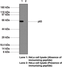
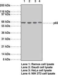
  • NF-κB p65 is a ubiquitously expressed transcription factor that is a subunit of the NF-κB complex and is encoded by the RELA gene in humans.{53059} It is composed of an N-terminal Rel homology domain, which contains the nuclear localization signal (NLS), and mediates dimerization, nuclear localization, and DNA and protein interactions, and two C-terminal transactivation domains that are subject to a variety of post-translational modifications and regulate the transcriptional activity of p65.{53059,53060} NF-κB p65 regulates the expression of a large number of genes in response to inflammatory and environmental cues that play critical roles in innate and adaptive immunity and cellular differentiation.{53060} Silencing of Rela induces tumor cell apoptosis in a murine Lewis lung carcinoma model, and RELA silencing in THP-1 monocytes decreases secreted levels of IL-1β and TNF-α induced by LPS.{53061,53062} Genome-wide deletion of Rela in mice is embryonic lethal.{5321} NF-κB p65 is overexpressed in the inflamed joints of patients with rheumatoid arthritis, and naïve CD4 T cells isolated from the whole blood of patients with multiple sclerosis have increased phosphorylation of NF-κB p65.{53065,53066} Cayman’s NF-κB (p65) Monoclonal Antibody – Biotinylated (Clone 112A1021) can be used for ELISA applications. The antibody recognizes NF-κB (p65) at 65 kDa from human, mouse, and rat samples.  


    Brand:
    Cayman
    SKU:13756 - 1 ea

    Available on backorder

  • NF-κB p65 is a ubiquitously expressed transcription factor that is a subunit of the NF-κB complex and is encoded by the RELA gene in humans.{53059} It is composed of an N-terminal Rel homology domain, which contains the nuclear localization signal (NLS), and mediates dimerization, nuclear localization, and DNA and protein interactions, and two C-terminal transactivation domains that are subject to a variety of post-translational modifications and regulate the transcriptional activity of p65.{53059,53060} NF-κB p65 regulates the expression of a large number of genes in response to inflammatory and environmental cues that play critical roles in innate and adaptive immunity and cellular differentiation.{53060} Silencing of Rela induces tumor cell apoptosis in a murine Lewis lung carcinoma model, and RELA silencing in THP-1 monocytes decreases secreted levels of IL-1β and TNF-α induced by LPS.{53061,53062} Genome-wide deletion of Rela in mice is embryonic lethal.{5321} NF-κB p65 is overexpressed in the inflamed joints of patients with rheumatoid arthritis, and naïve CD4 T cells isolated from the whole blood of patients with multiple sclerosis have increased phosphorylation of NF-κB p65.{53065,53066} Cayman’s NF-κB (p65) Monoclonal Antibody (Clone 112A1021) can be used for flow cytometry (FC), immunohistochemistry (IHC), and Western blot (WB) applications. The antibody recognizes NF-κB (p65) from human, mouse, and rat samples.  


    Brand:
    Cayman
    SKU:13752 - 1 ea

    Available on backorder

  • NF-κB p65 is a ubiquitously expressed transcription factor that is a subunit of the NF-κB complex and is encoded by the RELA gene in humans.{53059} It is composed of an N-terminal Rel homology domain, which contains the nuclear localization signal (NLS), and mediates dimerization, nuclear localization, and DNA and protein interactions, and two C-terminal transactivation domains that are subject to a variety of post-translational modifications and regulate the transcriptional activity of p65.{53059,53060} NF-κB p65 regulates the expression of a large number of genes in response to inflammatory and environmental cues that play critical roles in innate and adaptive immunity and cellular differentiation.{53060} Silencing of Rela induces tumor cell apoptosis in a murine Lewis lung carcinoma model, and RELA silencing in THP-1 monocytes decreases secreted levels of IL-1β and TNF-α induced by LPS.{53061,53062} Genome-wide deletion of Rela in mice is embryonic lethal.{5321} NF-κB p65 is overexpressed in the inflamed joints of patients with rheumatoid arthritis, and naïve CD4 T cells isolated from the whole blood of patients with multiple sclerosis have increased phosphorylation of NF-κB p65.{53065,53066} Cayman’s NF-κB (p65) NLS Polyclonal Antibody can be used for immunocytochemistry (ICC), immunofluorescence (IF), immunohistochemistry (IHC), IHC-paraffin (IHC-P), and Western blot (WB) applications. The antibody recognizes the NLS region of NF-κB to detect NF-κB at 65 kDa from human samples. It also recognizes the NLS region of NF-κB from chimpanzee, cow, gorilla, horse, monkey, and mouse samples.  


    Brand:
    Cayman
    SKU:13751 - 1 ea

    Available on backorder

  • NF-κB p65 is a ubiquitously expressed transcription factor that is a subunit of the NF-κB complex and is encoded by the RELA gene in humans.{53059} It is composed of an N-terminal Rel homology domain, which mediates dimerization, nuclear localization, and DNA and protein interactions, and two C-terminal transactivation domains that are subject to a variety of post-translational modifications and regulate the transcriptional activity of p65.{53059,53060} NF-κB p65 regulates the expression of a large number of genes in response to inflammatory and environmental cues that play critical roles in innate and adaptive immunity and cellular differentiation.{53060} Genome-wide deletion of Rela in mice is embryonic lethal.{5321} Silencing of NF-κB p65 induces tumor cell apoptosis in a murine Lewis lung carcinoma model and decreases secreted levels of IL-1β and TNF-α induced by LPS in THP-1 monocytes.{53061,53062} NF-κB p65 is overexpressed in inflamed joints of patients with rheumatoid arthritis, and naïve CD4 T cells isolated from the whole blood of patients with multiple sclerosis have increased phosphorylation of NF-κB p65.{53065,53066} Cayman’s NF-κB p65 (human, recombinant) protein can be used for Western blot and ELISA applications. Cayman’s NF-κB (p65) Polyclonal Antibody can be used for ELISA and Western blot applications. The antibody recognizes NF-κB at 65 kDa from human samples.  


    Brand:
    Cayman
    SKU:20671 - 100 µg

    Available on backorder

  • NF-κB p65 is a ubiquitously expressed transcription factor that is a subunit of the NF-κB complex and is encoded by the RELA gene in humans.{53059} It is composed of an N-terminal Rel homology domain, which contains the nuclear localization signal (NLS), and mediates dimerization, nuclear localization, and DNA and protein interactions, and two C-terminal transactivation domains that are subject to a variety of post-translational modifications and regulate the transcriptional activity of p65.{53059,53060} NF-κB p65 regulates the expression of a large number of genes in response to inflammatory and environmental cues that play critical roles in innate and adaptive immunity and cellular differentiation.{53060} Silencing of Rela induces tumor cell apoptosis in a murine Lewis lung carcinoma model, and RELA silencing in THP-1 monocytes decreases secreted levels of IL-1β and TNF-α induced by LPS.{53061,53062} Genome-wide deletion of Rela in mice is embryonic lethal.{5321} NF-κB p65 is overexpressed in the inflamed joints of patients with rheumatoid arthritis, and naïve CD4 T cells isolated from the whole blood of patients with multiple sclerosis have increased phosphorylation of NF-κB p65.{53065,53066} Cayman’s NF-ĸB (p65) Polyclonal Antibody (aa 2-17) can be used for Western blot (WB) applications. The antibody recognizes amino acids 2-17 of NF-κB (p65) at 60 kDa from human, chimpanzee, and monkey samples.  


    Brand:
    Cayman
    SKU:13757 - 1 ea

    Available on backorder

  • NF-κB p65 is a ubiquitously expressed transcription factor that is a subunit of the NF-κB complex and is encoded by the RELA gene in humans.{53059} It is composed of an N-terminal Rel homology domain, which contains the nuclear localization signal (NLS), and mediates dimerization, nuclear localization, and DNA and protein interactions, and two C-terminal transactivation domains that are subject to a variety of post-translational modifications and regulate the transcriptional activity of p65.{53059,53060} NF-κB p65 regulates the expression of a large number of genes in response to inflammatory and environmental cues that play critical roles in innate and adaptive immunity and cellular differentiation.{53060} Silencing of Rela induces tumor cell apoptosis in a murine Lewis lung carcinoma model, and RELA silencing in THP-1 monocytes decreases secreted levels of IL-1β and TNF-α induced by LPS.{53061,53062} Genome-wide deletion of Rela in mice is embryonic lethal.{5321} NF-κB p65 is overexpressed in the inflamed joints of patients with rheumatoid arthritis, and naïve CD4 T cells isolated from the whole blood of patients with multiple sclerosis have increased phosphorylation of NF-κB p65.{53065,53066} Cayman’s NF-κB (p65) Rabbit Monoclonal Antibody (Clone RM273) can be used for immunohistochemistry (IHC) and Western blot (WB) applications.  


    Brand:
    Cayman
    SKU:32223 - 100 µl

    Available on backorder

  • Cayman’s NF-κB (p65) Transcription Factor Assay is a non-radioactive, sensitive method for detecting specific transcription factor DNA binding activity in nuclear extracts and whole cell lysates. A 96-well enzyme-linked immunosorbent assay (ELISA) replaces the cumbersome radioactive electrophoretic mobility shift assay (EMSA). A specific double stranded DNA (dsDNA) sequence containing the NF-κB response element is immobilized to the wells of a 96-well plate. NF-κB contained in a nuclear extract, binds specifically to the NF-κB response element. NF-κB (p65) is detected by addition of specific primary antibody directed against NF-κB (p65). A secondary antibody conjugated to HRP is added to provide a sensitive colorimetric readout at 450 nm. Cayman’s NF-κB (p65) Transcription Factor Assay detects human NF-κB (p65). It will not cross-react with NF-κB (p50).  


    Brand:
    Cayman
    SKU:10007889 - 96 wells

    Available on backorder

  • Brand:
    Cayman
    SKU:780022 - 1 ea

    Available on backorder

  • The Cayman Chemical Nitrate/Nitrite Assay Kit provides an accurate and convenient method for measurement of total nitrate/nitrite concentration in a simple two-step process. The first step is the conversion of nitrate to nitrite utilizing nitrate reductase. The second step is the addition of the Griess Reagents which convert nitrite into a deep purple azo compound. Photometric measurement of the absorbance due to this azo chromophore accurately determines nitrite concentration. This kit can be used to measure nitrate and nitrite in plasma/serum, urine, tissue culture media, and tissue homogenates. However, it cannot be used to analyze nitrate and nitrite from an in vitro assay of nitric oxide synthase (NOS) in which excess NADPH has been added. For these assays a second method (LDH method) is utilized and is available from Cayman Chemical in a 96 well plate format (Item No. 760871).  


    Brand:
    Cayman
    SKU:780001 - 2 x 96 wells

    Available on backorder

  • NADPH is an essential cofactor for the function of the NOS enzyme. Unfortunately, NADPH interferes with the chemistry of the Griess reagents, which are the most commonly used reagents for nitrite detection. There are two ways to get around this limitation. Vanishingly small amounts of NADPH can be used, in conjunction with a catalytic system for recycling the spent NADP+ back to NADPH. This is the system used in Cayman’s first Nitrate/Nitrite Colorimetric Assay Kit (Item No. 780001). It works well for the analysis of fluids such as plasma and urine, and is also available in a high sensitivity fluorometric version (Item No. 780051) for the detection of low levels of nitrate and nitrite. However, this method cannot be used to analyze nitrate and nitrite from an in vitro NOS assay in which excess NADPH has been added. The second way to handle the NADPH interference problem is to go ahead and use an excess of NADPH. An additional step is added to the protocol to remove the excess NADPH. This is the avenue we have taken in the development of this nitrate/nitrite assay kit. This kit uses Lactate Dehydrogenase (LDH) to oxidize the excess NADPH. The kit is particularly well suited to measurements of NOS activity in vitro. High throughput screening applications using recombinant nitric oxide synthase (NOS) preparations can be quickly and accurately assayed with the LDH method kit. Each kit contains assay buffer, nitrate reductase, cofactors preparation, nitrate standard, lactate dehydrogenase, Griess Reagent R1, Griess Reagent R2, a 96 well plate, and complete instructions.  


    Brand:
    Cayman
    SKU:760871 - 96 wells

    Available on backorder

  • Nitric Oxide (NO) is produced in trace quantities by neurons, endothelial cells, platelets, and neutrophils in response to homeostatic stimuli. This NO is scavenged rapidly (t½ = 4 s) and acts in a paracrine fashion to transduce cellular signals. NO interacts with the HEME prosthetic group of guanylate cyclase, activating the enzyme and leading to increased cGMP levels. Other cells (macrophages, fibroblasts, and hepatocytes) also produce NO in micromolar concentrations in response to inflammatory or mitogenic stimuli. These very high levels of NO lead to the formation of peroxynitrite, destruction of iron-sulphur clusters, thiol nitrosation, and nitration of protein tyrosine residues. Thus, the amount of NO produced in different biological systems can vary over several orders of magnitude and its subsequent chemical reactivity is diverse. The final products of NO in vivo are nitrite (NO2-) and nitrate (NO3-). Relative proportions of NO2- and NO3- within a sample are variable and cannot be predicted with certainty. Therefore, the best index of total NO production is the sum of both of these products. Cayman Chemical’s Nitrate/Nitrite Fluorometric Assay Kit provides a convenient method for the quantitation of low levels of NO2- and NO3- in biological samples (particularly tissue culture medium). Previously undetectable concentrations of nitrate/nitrite in a variety of samples can be accurately quantitated using this kit as it extends the lower limit of detection by 20-fold over our popular colorimetric version. The minimum detectable quantity of NO2/NO3 is ~50 nM.  


    Brand:
    Cayman
    SKU:780051 - 2 x 96 wells

    Available on backorder

  • Nitrotyrosine is a post-translational modification that is formed by the nitration of tyrosine.{60009} Under conditions of oxidative stress, tyrosine is oxidized by reactive oxygen species (ROS) or, in the presence of hydrogen peroxide and nitrite, by myeloperoxidase (MPO) to yield a tyrosine radical that reacts with reactive nitrogen species (RNS), such as nitric oxide or peroxynitrite, to form nitrotyrosine. It exists in a free or protein-bound form and is commonly used as a marker of nitrosative or oxidative stress.{31710} Nitrotyrosine residues have been found in a variety of proteins, including LDL, surfactant protein A, angiotensin II, and human and bovine serum albumin.{60010} Free nitrotyrosine production induced by peroxynitrite is inhibited by the polyphenols epicatechin gallate, gallic acid, catechin, or epicatechin in cell-free assays, as well as in aortic rings isolated from normotensive and spontaneously hypertensive rats administered the antioxidant N-acetyl-cysteine (NAC; Item No. 20261).{60011,60012} Nitrotyrosine levels are increased in the affected tissues of numerous pathological conditions, including atherosclerosis, cancer, ulcerative colitis, Alzheimer’s disease, and Parkinson’s disease.{60010} Autoantibodies that recognize nitrotyrosinated proteins are increased in the synovium of patients with rheumatoid arthritis and are positively correlated with joint and tendon inflammation.{60009} Cayman’s Nitrotyrosine (Peptide) Polyclonal Antibody has been prepared by affinity purification to exclude non-nitrotyrosine antibodies generated against the antigenic peptide backbone. This antibody can be used for Western blot (WB) applications.  


    Brand:
    Cayman
    SKU:10006778 - 500 µl

    Available on backorder

  • Nitrotyrosine is a post-translational modification that is formed by the nitration of tyrosine.{60009} Under conditions of oxidative stress, tyrosine is oxidized by reactive oxygen species (ROS) or, in the presence of hydrogen peroxide and nitrite, by myeloperoxidase (MPO) to yield a tyrosine radical that reacts with reactive nitrogen species (RNS), such as nitric oxide or peroxynitrite, to form nitrotyrosine. It exists in a free or protein-bound form and is commonly used as a marker of nitrosative or oxidative stress.{31710} Nitrotyrosine residues have been found in a variety of proteins, including LDL, surfactant protein A, angiotensin II, and human and bovine serum albumin.{60010} Free nitrotyrosine production induced by peroxynitrite is inhibited by the polyphenols epicatechin gallate, gallic acid, catechin, or epicatechin in cell-free assays, as well as in aortic rings isolated from normotensive and spontaneously hypertensive rats administered the antioxidant N-acetyl-cysteine (NAC; Item No. 20261).{60011,60012} Nitrotyrosine levels are increased in the affected tissues of numerous pathological conditions, including atherosclerosis, cancer, ulcerative colitis, Alzheimer’s disease, and Parkinson’s disease.{60010} Autoantibodies that recognize nitrotyrosinated proteins are increased in the synovium of patients with rheumatoid arthritis and are positively correlated with joint and tendon inflammation.{60009} Cayman’s Nitrotyrosine Monoclonal Antibody can be used for ELISA and Western blot (WB) applications.  


    Brand:
    Cayman
    SKU:189542 - 200 µg

    Available on backorder

  • Nitrotyrosine is a post-translational modification that is formed by the nitration of tyrosine.{60009} Under conditions of oxidative stress, tyrosine is oxidized by reactive oxygen species (ROS) or, in the presence of hydrogen peroxide and nitrite, by myeloperoxidase (MPO) to yield a tyrosine radical that reacts with reactive nitrogen species (RNS), such as nitric oxide or peroxynitrite, to form nitrotyrosine. It exists in a free or protein-bound form and is commonly used as a marker of nitrosative or oxidative stress.{31710} Nitrotyrosine residues have been found in a variety of proteins, including LDL, surfactant protein A, angiotensin II, and human and bovine serum albumin.{60010} Free nitrotyrosine production induced by peroxynitrite is inhibited by the polyphenols epicatechin gallate, gallic acid, catechin, or epicatechin in cell-free assays, as well as in aortic rings isolated from normotensive and spontaneously hypertensive rats administered the antioxidant N-acetyl-cysteine (NAC; Item No. 20261).{60011,60012} Nitrotyrosine levels are increased in the affected tissues of numerous pathological conditions, including atherosclerosis, cancer, ulcerative colitis, Alzheimer’s disease, and Parkinson’s disease.{60010} Autoantibodies that recognize nitrotyrosinated proteins are increased in the synovium of patients with rheumatoid arthritis and are positively correlated with joint and tendon inflammation.{60009} Cayman’s Nitrotyrosine Monoclonal Antibody can be used for ELISA and Western blot (WB) applications.  


    Brand:
    Cayman
    SKU:189542 - 50 µg

    Available on backorder

  • Nitrotyrosine is a post-translational modification that is formed by the nitration of tyrosine.{60009} Under conditions of oxidative stress, tyrosine is oxidized by reactive oxygen species (ROS) or, in the presence of hydrogen peroxide and nitrite, by myeloperoxidase (MPO) to yield a tyrosine radical that reacts with reactive nitrogen species (RNS), such as nitric oxide or peroxynitrite, to form nitrotyrosine. It exists in a free or protein-bound form and is commonly used as a marker of nitrosative or oxidative stress.{31710} Nitrotyrosine residues have been found in a variety of proteins, including LDL, surfactant protein A, angiotensin II, and human and bovine serum albumin.{60010} Free nitrotyrosine production induced by peroxynitrite is inhibited by the polyphenols epicatechin gallate, gallic acid, catechin, or epicatechin in cell-free assays, as well as in aortic rings isolated from normotensive and spontaneously hypertensive rats administered the antioxidant N-acetyl-cysteine (NAC; Item No. 20261).{60011,60012} Nitrotyrosine levels are increased in the affected tissues of numerous pathological conditions, including atherosclerosis, cancer, ulcerative colitis, Alzheimer’s disease, and Parkinson’s disease.{60010} Autoantibodies that recognize nitrotyrosinated proteins are increased in the synovium of patients with rheumatoid arthritis and are positively correlated with joint and tendon inflammation.{60009} Cayman’s Nitrotyrosine Monoclonal Antibody – Biotinylated can be used for ELISA or Western blot (WB) applications.  


    Brand:
    Cayman
    SKU:10006966 - 100 µg

    Available on backorder

  • Nitrotyrosine is a post-translational modification that is formed by the nitration of tyrosine.{60009} Under conditions of oxidative stress, tyrosine is oxidized by reactive oxygen species (ROS) or, in the presence of hydrogen peroxide and nitrite, by myeloperoxidase (MPO) to yield a tyrosine radical that reacts with reactive nitrogen species (RNS), such as nitric oxide or peroxynitrite, to form nitrotyrosine. It exists in a free or protein-bound form and is commonly used as a marker of nitrosative or oxidative stress.{31710} Nitrotyrosine residues have been found in a variety of proteins, including LDL, surfactant protein A, angiotensin II, and human and bovine serum albumin.{60010} Free nitrotyrosine production induced by peroxynitrite is inhibited by the polyphenols epicatechin gallate, gallic acid, catechin, or epicatechin in cell-free assays, as well as in aortic rings isolated from normotensive and spontaneously hypertensive rats administered the antioxidant N-acetyl-cysteine (NAC; Item No. 20261).{60011,60012} Nitrotyrosine levels are increased in the affected tissues of numerous pathological conditions, including atherosclerosis, cancer, ulcerative colitis, Alzheimer’s disease, and Parkinson’s disease.{60010} Autoantibodies that recognize nitrotyrosinated proteins are increased in the synovium of patients with rheumatoid arthritis and are positively correlated with joint and tendon inflammation.{60009} Cayman’s Nitrotyrosine Polyclonal Antibody can be used for Western blot (WB) applications.  


    Brand:
    Cayman
    SKU:10189540 - 1 ea

    Available on backorder

  • The ion channels activated by glutamate are typically divided into two classes. Glutamate receptors that are activated by kainate and α-amino-3-hydroxy-5-methyl-4-isoxalone propionic acid (AMPA) are known as kainate/AMPA receptors (K/AMPAR). Those that are sensitive to Nmethyl- D-aspartate (NMDA) are designated NMDA receptors (NMDAR). The NMDAR plays an essential role in memory, neuronal development and it has also been implicated in several disorders of the central nervous system including Alzheimer’s, epilepsy and ischemic neuronal cell death. The NMDA receptor is also one of the principal molecular targets for alcohol in the CNS. The NMDAR is also potentiated by protein phosphorylation. The rat NMDAR1 (NR1) was the first subunit of the NMDAR to be cloned. The NR1 protein can form NMDA activated channels when expressed in Xenopus oocytes but the currents in such channels are much smaller than those seen in situ. Channels with more physiological characteristics are produced when the NR1-subunit is combined with one or more of the NMDAR2 (NR2 A-D) subunits.  


    Brand:
    Cayman
    SKU:10607 - 1 ea

    Available on backorder

  • The ion channels activated by glutamate are typically divided into two classes. Glutamate receptors that are activated by kainate and α-amino-3-hydroxy-5-methyl-4-isoxalone propionic acid (AMPA) are known as kainate/AMPA receptors (K/AMPAR). Those that are sensitive to N-methyl-Daspartate (NMDA) are designated NMDA receptors (NMDAR). The NMDAR plays an essential role in memory, neuronal development and it has also been implicated in several disorders of the central nervous system including Alzheimer’s, epilepsy and ischemic neuronal cell death. The NMDA receptor is also one of the principal molecular targets for alcohol in the CNS. The NMDAR is also potentiated by protein phosphorylation. The rat NMDAR1 (NR1) was the first subunit of the NMDAR to be cloned. The NR1 protein can form NMDA activated channels when expressed in Xenopus oocytes but the currents in such channels are much smaller than those seen in situ. Channels with more physiological characteristics are produced when the NR1 subunit is combined with one or more of the NMDAR2 (NR2 A-D) subunits.  


    Brand:
    Cayman
    SKU:10608 - 1 ea

    Available on backorder

  • The NMDA receptor (NMDAR) plays an essential role in memory, neuronal development, and it has also been implicated in several disorders of the central nervous system including Alzheimer’s disease, epilepsy, and ischemic neuronal death. {14637,14639,14638} The NR1 protein can form NMDA activated channels when expressed in Xenopus oocytes but the currents in such channels are much smaller than those seen in situ. Channels with more physiological characteristics are produced when the NR1 subunit is combined with one or more of the NMDAR2 (NR2 A-D) subunits. {14636} Phosphorylation of Tyr1252 in NR2B is thought to potentiate NMDA receptor-dependent influx of calcium.{14640}  


    Brand:
    Cayman
    SKU:10009759 - 1 ea

    Available on backorder

  • The NMDA receptor (NMDAR) plays an essential role in memory, neuronal development, and it has also been implicated in several disorders of the central nervous system including Alzheimer’s disease, epilepsy, and ischemic neuronal death. {14637,14639,14638} The NR1 protein can form NMDA activated channels when expressed in Xenopus oocytes but the currents in such channels are much smaller than those seen in situ. Channels with more physiological characteristics are produced when the NR1 subunit is combined with one or more of the NMDAR2 (NR2 A-D) subunits. {14636} Phosphorylation of Tyr1336 in NR2B is thought to potentiate NMDA receptor-dependent influx of calcium.{14640} Ischemia may also increase the phosphorylation of this site.  


    Brand:
    Cayman
    SKU:10009760 - 1 ea

    Available on backorder

  • The NMDA receptor (NMDAR) plays an essential role in memory, neuronal development and it has also been implicated in several disorders of the central nervous system including Alzheimer’s disease, epilepsy and ischemic neuronal death. {14637,14639,14638} The NMDA receptor is also one of the principal molecular targets for alcohol in the CNS. {14643,14633,14645} Channels with physiological characteristics are produced when the NR1 subunit is combined with one or more of the NMDAR2 (NR2 A-D) subunits. {14636} Overexpression of the NR2B-subunit of the NMDA Receptor has been associated with increases in learning and memory while aged, memory impaired animals have deficiencies in NR2B expression. {14635,14641} Recent work suggests that phosphorylation of Tyr1472 on NR2B may regulate the functional expression of the receptor in LTP and other forms of plasticity. {14634,14644}  


    Brand:
    Cayman
    SKU:10009761 - 1 ea

    Available on backorder

  • The ion channels activated by glutamate that are sensitive to N-methyl-D-aspartate (NMDA) are designated NMDA receptors (NMDAR). The NMDAR plays an essential role in memory, neuronal development and it has also been implicated in several disorders of the central nervous system including Alzheimer’s, epilepsy. and ischemic neuronal cell death. The NMDA receptor is also one of the principal molecular targets for alcohol in the CNS. The rat NMDAR1 (NR1) was the first subunit of the NMDAR to be clonedand it can form NMDA activated channels when expressed in Xenopus oocytes but the currents in such channels are much smaller than those seen in situ. Channels with more physiological characteristics are produced when the NR1 subunit is combined with one or more of the NMDAR2 (NR2 A-D) subunits. Overexpression of the NR2B-subunit of the NMDA receptor has been associated with increases in learning and memory while aged, memory impaired animals have deficiencies in NR2B expression. The NMDAR is also potentiated by protein phosphorylation.  


    Brand:
    Cayman
    SKU:10609 - 1 ea

    Available on backorder

  • Nitric oxide synthase (NOS) catalyzes the oxidation of arginine to nitric oxide (NO) and citrulline. Three distinct isoforms of NOS have been described having nomenclature based on the tissue source from which they were originally cloned. These three isoforms are neuronal/brain NOS (nNOS/bNOS/NOS-I), inducible NOS (iNOS/NOS-II), and endothelial NOS (eNOS/NOS-III).{4141,5917} nNOS is a soluble enzyme found in brain, the peripheral nervous system and skeletal muscle.{1280,4153} An alternately spliced form of nNOS (nNOSμ) containing a 34 amino acid insert has been identified in skeletal muscle.{2924} In neurons, protein-protein interactions with PSD95 and PSD93 via the PZD domain at the N-terminus of nNOS localizes the enzyme with NMDA receptors.{4287,6447} Although nNOS was originally thought to be constitutively expressed, abundant evidence suggests expression is regulated by a variety of conditions.{7458}  


    Brand:
    Cayman
    SKU:160870 - 500 µl

    Available on backorder

  • Brand:
    Cayman
    SKU:760873 - 1 ea

    Available on backorder

  • Brand:
    Cayman
    SKU:760874 - 1 ea

    Available on backorder

  • Brand:
    Cayman
    SKU:780014 - 1 ea

    Available on backorder

  • Nod2 is a member of the apoptosis regulating protein family that has caspase recruitment-domains (CARDs) and also includes Apaf-1 and Nod1.{8852} Nod2 contains two N-terminal CARDs, a nucleotide binding domain (NBD), and multiple C-terminal leucine-rich repeats (LRRs). Nod2 is expressed in monocytes whereas Nod1 (CARD4) is expressed in multiple tissues.{8852,12079,8407,12078} Both Nod1 and Nod2 act as intracellular receptors for bacterial lipopolysaccharides, activate NF-kB, and contribute to inflammatory bowel disease.{8407,8408,8855,8859} A frameshift mutation in the NOD2 gene is associated with susceptibility to Crohn’s disease, possibly by causing truncation of the tenth LRR.{8853,8854} Cayman Chemical’s NOD2 Monoclonal Antibody can be used for Western blot and immunohistochemical analysis of Nod2 on samples of human origin. The antibody will also recognize three Nod2 polymorphisms: L1007finsC, G908R, and R702W.{12079}  


    Brand:
    Cayman
    SKU:10004942 - 1 ea

    Available on backorder

  • Nod2 is a member of a protein family of apoptosis regulators which includes Apaf-1 and Nod1/CARD4.{8852} Nod2 contains two N-terminal caspase recruitment domains (CARDs), a nucleotide binding domain (NBD) and C-terminal leucine rich repeats (LRRs). Nod1/CARD4 is broadly expressed in tissues; however, Nod2 expression is limited to monocytes.{8852,8407} Both Nod1 and Nod2 activate NF-kB and act as intracellular receptors for bacterial lipopolysaccharides (LPS).{8407,8859,8855,8408} The LRRs of both Nod1 and Nod2 are essential for LPS-induced activation of NF-kB.{8855} A frameshift mutation in the NOD2 gene confers susceptibility to Crohn’s disease, possibly by causing truncation of the 10th LRR resulting in a protein that is unresponsive to LPS.{8853,8854}  


    Brand:
    Cayman
    SKU:160777 - 1 ea

    Available on backorder

  • Brand:
    Cayman
    SKU:600009 - 500 µl

    Available on backorder

  • Brand:
    Cayman
    SKU:600009 - 6 ml

    Available on backorder

  • Present in nature in the form of iodide and iodate, iodine is a solid halogen at normal temperature. It is used in medicine, in the pharmaceutical and food industry. Food is the principal daily supply of iodide in human body.{35362} Iodide is important in basal metabolism and permits temperature regulation, intellectual development for children, muscular development, normal heart function and growth of skeleton. Iodide transport is the basis for an emerging approach of selective cancer cell destruction.{35363,35364} Iodide uptake from blood into thyroid follicular cells is the first step in the biosynthesis of thyroid hormones T4 and T3, known to regulate many essential biological processes.{35365,35366}Thyroid hormones are indispensable for body development. This transport is mediated by NIS (sodium iodide symporter), an intrinsic membrane glycoprotein located in the basolateral membrane of thyrocytes. Since the discovery of NIS, thorough biochemical analysis has elucidated the mechanism of basolateral iodide transport and revealed the key role of NIS in thyroid diseases such as thyroid cancer, autoimmune disease, and congenital hypothyroidism.{35367} If rate is not in the normal proportion, some diseases can be developed as underactive thyroid if the rate is too down or overactive thyroid if the rate is too up. Other diseases exist as chronic thyroiditis of Hashimoto or cancer of the thyroid gland.{35368} Iodide deficiency is at origins of many thyroid metabolism disorders, this is why it is important to control rate of iodide to prevent all of these diseases. This Iodide Assay is based on the oxido-reduction reaction: cerium(IV) is reduced by arsenic(III). The reduction of yellow (420 nm) cerium(IV) to colorless cerium(III) by arsenic(III) proceeds very slowly but traces of iodide strongly accelerate this reaction with the rate being directly proportional to iodide concentration. For a given time, decay is inversely proportional to iodide concentration in well. This method is simple and nonradioactive, and as such it can be used widely.[Bertin Catalog No. D05076.96]  


    Brand:
    Cayman
    SKU:25659 - 96 wells

    Available on backorder

  • The NOS activity assay kit measures NOS activity by monitoring the conversion of radiolabeled arginine to citrulline. This assay is simple, sensitive, and specific for NOS activity and can be used with both crude and purified enzyme preparations. The kit includes sufficient materials and reagents for 50 total reactions. Radiolabeled arginine and NADPH are not included with the kit.  


    Brand:
    Cayman
    SKU:781001 - 1 ea

    Available on backorder

  • Nuclear factor erythroid 2-related factor 2 (Nrf2) is a basic leucine zipper transcription factor encoded by NFE2L2 in humans that regulates the cellular antioxidant response.{53865} It is a 605-amino acid protein comprised of seven highly conserved Nrf2-ECH (Neh) homology domains.{53866,53867} Nrf2 contains a large central domain that recruits transcriptional co-activators, mediates heterodimerization with the transcription factor Maf, and can be phosphorylated by GSK3 or the ubiquitin ligase adapter protein β-TrCP.{53868,53867} The Nrf2 central domain is flanked by an N-terminal Neh2 domain that binds the negative regulator Kelch-like ECH-associated protein 1 (Keap1; Item No. 32035) and a C-terminal Neh3 domain that is required for transactivation.{53867} Nrf2 is ubiquitously expressed and, under homeostatic conditions, associates with Keap1 in the cytoplasm, preventing Nrf2 nuclear translocation and promoting its ubiquitination and proteasomal degradation.{53866,53869} In the presence of xenobiotic electrophiles or oxidants, Keap1 releases Nrf2 which translocates to the nucleus, dimerizes with Maf, and induces the expression of a variety of cytoprotective genes that have antioxidant, anti-inflammatory, and metabolic functions.{53870,53745,53865} Nrf2 siRNA knockdown increases DNA-crosslinking and apoptosis induced by cisplatin (Item No. 13119) in A549 cancer cells, indicating that Nrf2 contributes to chemoresistance.{53871} Tumor Nrf2 levels are increased in patients with non-small cell lung cancer (NSCLC) and this increase is associated with poor overall survival.{53872} Cayman’s Nrf2 (C-Term) Polyclonal Antibody can be used for flow cytometry (FC), immunocytochemistry (ICC), immunofluorescence (IF), and Western blot (WB) applications. The antibody preferentially recognizes polyubiquitinated Nrf2 at 90 kDa from human, mouse, and rat samples.  


    Brand:
    Cayman
    SKU:10214 - 1 ea

    Available on backorder

  • Nuclear factor erythroid 2-related factor 2 (Nrf2) is a basic leucine zipper transcription factor encoded by NFE2L2 in humans that regulates the cellular antioxidant response.{53865} It is a 605-amino acid protein comprised of seven highly conserved Nrf2-ECH (Neh) homology domains.{53866,53867} Nrf2 contains a large central domain that recruits transcriptional co-activators, mediates heterodimerization with the transcription factor Maf, and can be phosphorylated by GSK3 or the ubiquitin ligase adapter protein β-TrCP.{53867,53868} The Nrf2 central domain is flanked by an N-terminal Neh2 domain that binds the negative regulator Kelch-like ECH-associated protein 1 (Keap1; Item No. 32035) and a C-terminal Neh3 domain that is required for transactivation.{53867} Nrf2 is ubiquitously expressed and, under homeostatic conditions, associates with Keap1 in the cytoplasm, preventing Nrf2 nuclear translocation and promoting its ubiquitination and proteasomal degradation.{53866,53869} In the presence of xenobiotic electrophiles or oxidants, Keap1 releases Nrf2 which translocates to the nucleus, dimerizes with Maf, and induces the expression of a variety of cytoprotective genes that have antioxidant, anti-inflammatory, and metabolic functions.{53865,53870,53745} Nrf2 siRNA knockdown increases DNA-crosslinking and apoptosis induced by cisplatin (Item No. 13119) in A549 cancer cells, indicating that Nrf2 contributes to chemoresistance.{53871} Tumor Nrf2 levels are increased in patients with non-small cell lung cancer (NSCLC) and this increase is associated with poor overall survival.{53872} Cayman’s Nrf2 (N-Term) Polyclonal Antibody can be used for flow cytometry (FC), immunocytochemistry (ICC), and Western blot (WB) applications. The antibody recognizes native and polyubiquitinated Nrf2 at 67 and 90 kDa, respectively, from human, mouse, and rat samples.  


    Brand:
    Cayman
    SKU:14114 - 1 ea

    Available on backorder

  • Nuclear factor E2-related factor 2 (Nrf2) is a transcription factor that plays a key role in maintaining redox homeostasis via its interaction with a cysteine-rich protein Kelch-like ECH-associated protein 1 (Keap1). In resting cells, Nrf2 and Keap1 form a tight complex, which is targeted for degradation by proteasomes. Under oxidative stress, Nrf2 is released from the Nrf2/Keap1 complex and translocates to the nucleus where it is able to induce the expression of a battery of genes encoding diverse cytoprotective proteins, including antioxidative enzymes, anti-inflammatory mediators, and proteasomes. Cayman’s Nrf2 Transcription Factor Assay is a non-radioactive, colorimetric method for detecting specific transcription factor DNA binding activity in nuclear extracts. A specific double stranded DNA (dsDNA) sequence containing the Nrf2 response element is immobilized onto the wells of a 96-well plate. Nrf2 contained in nuclear extract samples will bind specifically to the Nrf2 response element and can be detected by addition of a specific Nrf2 antibody and a secondary antibody conjugated to HRP.  


    Brand:
    Cayman
    SKU:600590 - 96 wells

    Available on backorder

  • Obestatin is an amidated peptide of 23 amino-acids with an alpha-helical secondary conformation. It was first isolated from rat stomach in 2005. Obestatin is a preproghrelin-derived peptide produced by many tissues or organs, including the stomach, pancreas, adipose tissue, skeletal muscle, and heart. Obestatin was originally identified as an anorexigenic peptide that  reduces food intake and body weight by opposing the actions of ghrelin. It has also been considered to be an antidiabetic peptide that positively influences glucose and lipid metabolism. It may also play a role in the regulation of blood pressure as its plasma concentration increases in hypertensive patients. Obestatin may also have a role in the regulation of anxiety and memory improvement. [Bertin Catalog No. A05036]  


    Brand:
    Cayman
    SKU:18902 - 96 wells

    Available on backorder

  • Obestatin is an amidated peptide of 23 amino acids with an α-helical secondary conformation. It was first isolated from rat stomach in 2005. Obestatin is a preproghrelin-derived peptide produced by many tissues or organs, including the stomach, pancreas, adipose tissue, skeletal muscle, and the heart. Obestatin was originally identified as an anorexigenic peptide that reduces food intake and body weight by opposing the actions of ghrelin. It has been considered to be an antidiabetic peptide that positively influences glucose and lipid metabolism. It may also play a role in the regulation of blood pressure as its plasma concentration increases in hypertensive patients. Obestatin may also have a role in the regulation of anxiety and memory improvement. [Bertin Catalog No. A05035]  


    Brand:
    Cayman
    SKU:18903 - 96 wells

    Available on backorder

  • Octamer-binding transcription factor 4 (Oct4) is an essential regulator of embryonic stem cell pluripotency and used as a marker for undifferentiated cells. It also plays a role in gene expression, DNA repair and/or replication, signal transduction and tumorigenesis. Cayman’s Oct4 Transcription Factor Assay is a non-radioactive, sensitive method for detecting specific transcription factor-DNA binding activity in nuclear extracts. A 96-well enzyme-linked immunosorbent assay (ELISA) replaces the cumbersome radioactive electrophoretic mobility shift assay (EMSA). A specific double-stranded DNA (dsDNA) sequence containing the Oct4 response element is immobilized onto the wells of a 96-well plate. Oct4, contained in a nuclear extract, binds specifically to the Oct4 response element. Oct4 is detected by the addition of a specific primary antibody directed against Oct4. A secondary antibody conjugated to HRP is added to provide a sensitive colorimetric readout at 450 nm.  


    Brand:
    Cayman
    SKU:601080 - 96 wells

    Available on backorder

  • Oligodendrocyte lineage transcription factor 1 (Olig1) is a basic helix-loop-helix transcription factor that promotes the formation and maturation of oligodendrocytes.{55117} It is expressed in the spinal cord and neuroepithelium of the ventral forebrain, colocalizing with the oligodendrocyte precursor markers Sox10 and Pdgfra. Olig1 is localized to the nucleus at the embryonic stage and is translocated to the cytoplasm after birth.{55118} However, Olig1 is translocated back to the nucleus of oligodendrocyte precursors following a demyelinating injury. Olig1-/- mice exhibit delayed, but not impaired, spinal cord oligodendrocyte differentiation and maturation, indicating that Olig1 is non-essential to oligodendrocyte differentiation.{55119} Olig1 knockdown delays mean disease onset and reduces disease severity in a mouse model of myelin oligodendrocyte glycoprotein-induced experimental autoimmune encephalomyelitis.{55120} Cayman’s Olig1 Polyclonal Antibody can be used for immunocytochemistry (ICC), immunohistochemistry (IHC), immunoprecipitation (IP), and Western blot (WB) applications. The antibody recognizes Olig1 at approximately 27 kDa from human, mouse, and rat samples.  


    Brand:
    Cayman
    SKU:29287 - 100 µl

    Available on backorder

  • Oligodendrocyte lineage transcription factor 2 (Olig2) is a basic helix-loop-helix transcription factor that is essential to the formation of oligodendrocytes and motor neurons in the spinal cord and somatic motor neurons in the hindbrain.{46965} It represses differentiation and sustains the replication-competent state of uncommitted progenitor cells to expand the progenitor cell pool during early development and promotes the cell fate decision to form oligodendrocyte progenitors and motor neurons in the spinal cord during late development. It also suppresses the formation of astrocytes in the spinal cord. Olig2 contains a triple serine motif at positions 10, 13, and 14 near its amino terminus that is subject to phosphorylation.{46966} Phosphorylation of Olig2 at this motif (phospho-Ser10,13,14) is developmentally regulated and decreases as Olig2+ progenitor cells mature into terminally differentiated oligodendrocytes. EB5 neurospheres expressing a triple phosphomimetic mutant of Olig2 induce more rapid tumor formation and decreased survival in mice compared with neurospheres expressing wild-type Olig2. Cayman’s Olig2 (Phospho-Ser10,13,14) Polyclonal Antibody can be used for Western blot (WB) applications. The antibody recognizes Olig2 (phospho-Ser10,13,14) at approximately 32 kDa from human, mouse, and rat samples.  


    Brand:
    Cayman
    SKU:29289 - 100 µl

    Available on backorder

  • Oligodendrocyte lineage transcription factor 2 (Olig2) is a basic helix-loop-helix transcription factor that is essential to the formation of oligodendrocytes and motor neurons in the spinal cord and somatic motor neurons in the hindbrain.{46965} It represses differentiation and sustains the replication competent state of uncommitted progenitor cells to expand the progenitor cell pool during early development and promotes the cell fate decision to form oligodendrocyte progenitors and motor neurons in the spinal cord during late development. It also suppresses the formation of astrocytes in the spinal cord. Olig2 overexpression enhances oligodendrocyte regeneration and myelin repair in a mouse model of lysophosphatidylcholine-induced demyelination.{56201} Knockdown of Olig2 in neurospheres prevents glioma formation but not neurosphere engraftment in mice.{56202} Cayman’s Olig2 Polyclonal Antibody can be used for immunocytochemistry (ICC), immunohistochemistry (IHC), immunoprecipitation (IP), and Western blot (WB) applications. The antibody recognizes Olig1 at approximately 32 kDa from human, mouse, and rat samples.  


    Brand:
    Cayman
    SKU:29288 - 100 µl

    Available on backorder

  • Optineurin (OPTN) is an adapter protein with roles in autophagy, vesicular trafficking, and inflammatory responses to viral and bacterial pathogens.{47745} OPTN binds to ubiquitinated cargo via its ubiquitin-binding domain and targets them for autophagic degradation by recruiting LC3-expressing phagophore membranes via its LC3-interacting region. It is phosphorylated by TANK binding kinase 1 (TBK1), which increases the binding affinity of OPTN for polyubiquitinated mitochondria, promotes TBK1 activation and recruitment to the mitochondria, and permits the degradation of damaged mitochondria via PINK1/parkin-dependent mitophagy.{47746} OPTN regulates transferrin receptor trafficking and recycling by mediating the association between the membrane trafficking protein Rab8 and its activator TBC1D17.{47747} Mutation of the OPTN ubiquitin binding domain reduces LPS- and poly(I:C)-induced increases in IFN-β levels in bone marrow-derived macrophages (BMDMs).{47748} OPTN knockout increases mortality and reduces plasma levels of TNF-α and IL-6 in a mouse model of E. coli-induced peritonitis.{47749} Mutations in OPTN are associated with primary open angle glaucoma, amyotrophic lateral sclerosis (ALS), Paget’s disease, and Alzheimer’s disease.{47745,47750} Cayman’s Optineurin Monoclonal Antibody (Clone 8E12) can be used for immunocytochemistry applications.  


    Brand:
    Cayman
    SKU:27230 - 100 µg

    Available on backorder

  • Optineurin, originally named FIP-2, is a 74 kDa protein implicated in signal transduction of the tumor necrosis factor (TNF) pathway as well as membrane traffic regulation and cellular morphogenesis. Optineurin protein is also involved in apoptosis, although the exact role is currently unknown. Increased apoptosis of trabecular meshwork cells is a factor in the pathogenesis of glaucoma and mutations in the optineurin gene have been correlated with the some types of the disease. Cayman Chemical is a leading provider of prostaglandin analogs, such as latanoprost and fluprostenol, currently utilized for lowering the intraocular pressure (IOP) associated with glaucoma. Our optineurin polyclonal antibody is another research tool useful for delineating the physiology of the disease.  


    Brand:
    Cayman
    SKU:100000 - 1 ea

    Available on backorder

  • Optineurin (originally named FIP-2) is a 74 kDa protein implicated in signal transduction of the tumor necrosis factor (TNF) pathway as well as membrane traffic regulation and cellular morphogenesis. Optineurin protein is also involved in apoptosis, although the exact role is currently unknown. Increased apoptosis of trabecular meshwork cells is a factor in the pathogenesis of glaucoma and mutations in the optineurin gene have been correlated with the some types of the disease. Cayman Chemical is a leading provider of prostaglandin analogs, such as latanoprost and fluprostenol, currently utilized for lowering the intraocular pressure (IOP) associated with glaucoma. Our optineurin polyclonal antibody is another research tool useful for delineating the physiology of the disease.  


    Brand:
    Cayman
    SKU:100002 - 500 µl

    Available on backorder

  • The Orexin 1 Receptor (OX1R) may be an important therapeutic target for treatment of sleep disorders, obesity, emotional stress, and addiction. Cayman’s OX1R Reporter Assay Kit consists of a 96-well plate coated with a transfection complex containing DNA constructs for OX1R, a recombinant G-protein, and a cAMP response element regulated Secreted Alkaline Phosphatase (SEAP) reporter (OX1R Reverse Transfection Strip Plate). Cells grown on the transfection complex will express OX1R at the cell surface and the recombinant G-protein inside the cell. Binding of agonists to OX1R initiates a signal transduction cascade resulting in expression of SEAP which is secreted into the cell culture medium. Aliquots of culture medium are removed at time intervals beginning at about 16 hours and SEAP activity is measured following addition of a luminescence-based alkaline phosphatase substrate provided in the kit. The kit is easy to use and can be easily adapted to high throughput screening for therapeutic compounds regulating activation of OX1R. A known OX1R agonist, Orexin A, is included in the kit for use as a positive control. The kit provides sufficient reagent to measure SEAP activity at three time points, using the black plates provided.  


    Brand:
    Cayman
    SKU:600240 - 100 tests

    Available on backorder

  • The Orexin 2 Receptor (OX2R) may be an important therapeutic target for treatment of sleep disorders, obesity, emotional stress, and addiction. Cayman’s OX2R Reporter Assay Kit consists of a 96-well plate coated with both OX2R and Secreted Alkaline Phosphatase (SEAP) reporter constructs (OX2R Reverse Transfection Strip Plate). Cells grown on the transfection complex will express OX2R at the cell surface and the recombinant G-protein inside the cell. Binding of agonists to OX2R initiates a signal transduction cascade resulting in expression of SEAP which is secreted into the cell culture medium. Aliquots of culture medium are removed at time intervals beginning at about 16 hours and SEAP activity is measured following addition of a luminescence-based alkaline phosphatases substrate provided in the kit. The kit is easy to use and can be easily adapted to high throughput screening for therapeutic compounds regulating activation of OX2R. A known OX2R agonist, Orexin A, is included in the kit for use as a positive control. The kit provides sufficient reagent to measure SEAP activity at three time points, using the black plates provided.  


    Brand:
    Cayman
    SKU:600250 - 100 tests

    Available on backorder

  • The oxidation of low density lipoprotein is thought to be one of the first steps in atherosclerosis. The uptake of oxidized LDL (oxLDL) by macrophages activates pro-inflammatory mediators that promote disease progression. Cayman’s Oxidized LDL Uptake Assay Kit is a convenient tool to study modulators of cellular uptake of oxLDL. The kit employs a fluorescently tagged, human oxLDL along with myricetin, an inhibitor of uptake that reduces CD36 expression.{16736} The reagents provided in this kit are sufficient to run a single plate of cultured cells by either flow cytometry or fluorescence microscopy.  


    Brand:
    Cayman
    SKU:601180 - 1 ea

    Available on backorder

  • Cayman’s Oxycodone ELISA Kit is a competitive assay which can be used to meausre oxycodone in urine, serum, and plasma. The standard curve spans the range of 3.9-500 pg/ml.  


    Brand:
    Cayman
    SKU:501590 - 96 solid wells

    Available on backorder

  • Cayman’s Oxycodone ELISA Kit is a competitive assay which can be used to meausre oxycodone in urine, serum, and plasma. The standard curve spans the range of 3.9-500 pg/ml.  


    Brand:
    Cayman
    SKU:501590 - 96 strip wells

    Available on backorder