ELISA Kits
Showing 2701–2850 of 3623 results
-
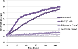
The oxygen consumption rate (OCR) of cells is an important indicator of normal cellular function. It is used as a parameter to study mitochondrial function as well as a marker of factors triggering the switch from healthy oxidative phosphorylation to aerobic glycolysis in cancer cells. Oxygen consumption is traditionally measured by a cumbersome oxygen electrode, a specialized piece of equipment that typically yields low sample throughput. A Phosphorescent Oxygen Probe has proven to be useful in measuring oxygen consumption rates in whole cells. Cayman’s cell-based Oxygen Consumption Rate Assay Kit utilizes this newly developed phosphorescent oxygen probe to measure oxygen consumption rate in living cells. Antimycin A, an inhibitor of the mitochondrial electron transport chain, is included to be used as a positive control. Glucose oxidase is also included in the kit to be used as a reference for oxygen depletion. The kit is easy to use and can be easily adapted to high throughput screening for compounds which modulate oxygen consumption rate.
Brand:CaymanSKU:600800 - 96 wellsAvailable on backorder
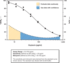
Oxytocin is a hypothalamic peptide hormone that is stored in the posterior pituitary gland. It is released into the bloodstream during parturition and lactation and is also involved in other social and sexual behaviors, neuropsychiatric disorders, and the maintenance of water and sodium homeostasis. Cayman’s Oxytocin ELISA Kit is a competitive assay that can be used for quantification of oxytocin in plasma. The assay has a range from 5.9-750 pg/ml and a sensitivity (80% B/B0) of approximately 20 pg/ml.
Brand:CaymanSKU:500440 - 480 solid wellsAvailable on backorder
Oxytocin is a hypothalamic peptide hormone that is stored in the posterior pituitary gland. It is released into the bloodstream during parturition and lactation and is also involved in other social and sexual behaviors, neuropsychiatric disorders, and the maintenance of water and sodium homeostasis. Cayman’s Oxytocin ELISA Kit is a competitive assay that can be used for quantification of oxytocin in plasma. The assay has a range from 5.9-750 pg/ml and a sensitivity (80% B/B0) of approximately 20 pg/ml.
Brand:CaymanSKU:500440 - 480 strip wellsAvailable on backorder
Oxytocin is a hypothalamic peptide hormone that is stored in the posterior pituitary gland. It is released into the bloodstream during parturition and lactation and is also involved in other social and sexual behaviors, neuropsychiatric disorders, and the maintenance of water and sodium homeostasis. Cayman’s Oxytocin ELISA Kit is a competitive assay that can be used for quantification of oxytocin in plasma. The assay has a range from 5.9-750 pg/ml and a sensitivity (80% B/B0) of approximately 20 pg/ml.
Brand:CaymanSKU:500440 - 96 solid wellsAvailable on backorder
Oxytocin is a hypothalamic peptide hormone that is stored in the posterior pituitary gland. It is released into the bloodstream during parturition and lactation and is also involved in other social and sexual behaviors, neuropsychiatric disorders, and the maintenance of water and sodium homeostasis. Cayman’s Oxytocin ELISA Kit is a competitive assay that can be used for quantification of oxytocin in plasma. The assay has a range from 5.9-750 pg/ml and a sensitivity (80% B/B0) of approximately 20 pg/ml.
Brand:CaymanSKU:500440 - 96 strip wellsAvailable on backorder
Brand:CaymanSKU:400444 - 7.5 ngAvailable on backorder
The three mitogen-activated protein kinases (MAPKs) are evolutionarily conserved protein kinases that control a vast array of cellular processes. p38 MAPK is one of these kinases and it is activated by both inflammatory cytokines and by stress.{14317,14326} The p38 MAPK is thought to be particularly important in diseases like asthma and autoimmunity but it also plays important roles in the stress response of the nervous system.{14318,14325} Like the other MAPKs, p38 is activated by a dual specificity kinase that phosphorylates Thr180 and Tyr182.{14316}
Brand:CaymanSKU:10009177 - 1 eaAvailable on backorder
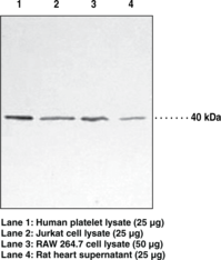
p38 mitogen-activated protein kinase (MAPK) is a member of the serine-threonine MAPK family that triggers many cellular processes including cell cycle, development, and apoptosis.{15420,15421} These kinases are activated by environmental stress signals such as osmotic shock, infection, and cytokines causing phosphorylation of p38 MAPK. This results in a phosphorylation cascade, activating transcription factors, and inducing gene expression.{15420,15424} p38 MAPK is widely expressed in heart, brain, skeletal muscle, platelets, and immune cells. Due to this distribution, p38 MAPK plays a role in cardiovascular disease, arthritis, and cancer.{15421,15422,1523,15424} It is mainly present in the cytosol, but can also be found in the nucleus after activation.{15421} Based on the amino acid sequence, the expected molecular weight of this protein is 41 kDa.
Brand:CaymanSKU:10011301 - 1 eaAvailable on backorder
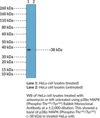
p38 MAPK is a serine/threonine protein kinase and member of the MAPK family with roles in the regulation of immune responses and embryonic development, as well as cell differentiation, metabolism, and survival.{60066,60067} It exists as 4 isoforms, p38α, -β, -γ, and -δ, encoded by MAPK14, MAPK11, MAPK12, and MAPK13, respectively, in humans. p38α MAPK is ubiquitously expressed, with the highest levels of expression in the heart, skeletal muscle, and brain.{60066,60068} It is activated via dual phosphorylation of threonine 180 (Thr180) and tyrosine 182 (Tyr182) by the MAP2K kinases MKK3 and MKK6 in response to LPS or the production of inflammatory cytokines and induces signaling through protein kinases, transcription factors, and transcriptional regulators, among others.{60066,60067} Levels of activated p38α MAPK (p38α phospho-Thr180/Tyr182) are increased and positively correlated with apoptosis in DU145 and PC3 prostate cancer cells in response to cisplatin (Item No. 13119).{60070} p38α Phospho-Thr180/Tyr182 levels are also increased in adult rat ventricular monocytes during stimulated ischemia.{60071} Cayman’s p38α MAPK (Phospho-Thr180/Tyr182) Rabbit Monoclonal Antibody can be used for immunohistochemistry (IHC) and Western blot (WB) applications. The antibody recognizes p38α MAPK (phospho-Thr180/Tyr182) at approximately 38 kDa from human samples.
Brand:CaymanSKU:32197 - 100 µlAvailable on backorder
p38 MAPK is a serine/threonine protein kinase and member of the MAPK family with roles in the regulation of immune responses and embryonic development, as well as cell differentiation, metabolism, and survival.{60066,60067} It exists as 4 isoforms, p38α, -β, -γ, and -δ, encoded by MAPK14, MAPK11, MAPK12, and MAPK13, respectively, in humans. p38α MAPK is ubiquitously expressed, with the highest levels of expression in heart, skeletal muscle, and brain.{60066,60068} It is activated via dual phosphorylation of threonine 180 and tyrosine 182 by the MAP2K kinases MKK3 and MKK6 in response to LPS or the production of inflammatory cytokines.{60066,60067} Downstream signaling targets of p38α MAPK include protein kinases, transcription factors, and transcriptional regulators, among others.{60067} Knockdown of Mapk14 is embryonic lethal, while macrophage-specific deletion of Mapk14 inhibits inflammatory cytokine production and is protective against cecal ligation and puncture-induced sepsis in mice.{60067,60068} Mapk14 knockdown also increases lysosomal degradation of β-secretase 1 (BACE1) and decreases amyloid-β (Aβ) production in the APP/PS1 double transgenic mouse model of Alzheimer’s disease.{60069} Cayman’s p38α MAPK Monoclonal Antibody (Clone RM245) can be used for immunohistochemistry (IHC) and Western blot (WB) applications. The antibody recognizes p38α MAPK at approximately 38 kDa from human samples.
Brand:CaymanSKU:32199 - 100 µlAvailable on backorder
This antibody was purified by sequential peptide-affinity chromatography to select only for IgG specific for p53 (Ser392). Cellular p53, often called the ‘guardian of the genome’, is a transcription factor that is activated in response to cellular stress (DNA damage, hypoxia, heat shock, etc.) and acts to prevent further proliferation of the stressed cell by induction of cell cycle arrest or apoptotic mediators.{8237} Nearly 50% of human tumors have mutated or non-functional p53. p53 amino acid residues can be modified by phosphorylation and acetylation. In vivo phosphorylation of p53 residues alters signal transduction that warrant further investigation.{11275,11834}
Brand:CaymanSKU:10004807 - 1 eaAvailable on backorder
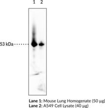
Epitope: Binds to N-terminal amino acids 16-25 of wild-type and mutant p53 Cellular p53, often called the ‘guardian of the genome,’ is a transcription factor that is activated in response to cellular stress (DNA damage, hypoxia, heat shock, etc.) and acts to prevent further proliferation of the stressed cell by induction of cell cycle arrest or apoptotic mediators.{8237} Nearly 50% of human tumors have mutated or non-functional p53. This antibody is suitable for a variety of techniques to distinguish between p53 positive and p53 negative tumors.{11929} This antibody binds the N-terminal (amino acids 16-25) of wild-type and mutant p53. This antibody does not work with frozen tissue sections. Cayman’s p53 Monoclonal Antibody (Clone BP53-12) can be used for flow cytometry, immunohistochemistry, immunoprecipitation, and Western blot applications. The antibody recognizes p53 at 53 kDa from human and mouse samples.
Brand:CaymanSKU:10004806 - 1 eaAvailable on backorder
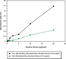
The tumor suppressor protein p53 plays a crucial role in coordinating cellular responses to genotoxic stress and holds many important clinical implications in the treatment of cancer. Cayman’s p53 Transcription Factor Assay is a non-radioactive, sensitive method for detecting specific transcription factor DNA binding activity in nuclear extracts. A specific double-stranded DNA (dsDNA) sequence containing the p53 response element is immobilized onto the wells of a 96-well plate. p53 contained in a nuclear extract, binds specifically to the p53 response element and is detected by addition of a specific primary antibody directed against p53. A secondary antibody conjugated to HRP is added to provide a sensitive colorimetric readout at 450 nm.
Brand:CaymanSKU:600020 - 96 wellsAvailable on backorder
p62, also known as sequestosome-1 (SQSTM1), is a 62 kDa protein that acts as a signaling hub and autophagy substrate and adaptor.{39547,39548} It is a multi-domain protein that includes a Phox1 and Bem1p (PB1) domain, a zinc finger, a tumor necrosis factor receptor-associated factor 6 (TRAF6) binding domain, a ubiquitin-associated (UBA) domain, LC3- and Keap1-interacting regions, as well as two nuclear localization and one nuclear export sequence. p62/SQSTM1 is constitutively expressed and is primarily localized in the cytoplasm, however, it is also expressed in the nucleus, autophagosomes, lysosomes, and inclusion bodies containing polyubiquitinated protein aggregates. It is overexpressed in a variety of human cancer cells as well as in the chronic liver diseases alcoholic and non-alcoholic steatohepatitis (NASH). p62/SQSTM1 binds to ERK1, RIP1, TRAF6, Raptor, PKC, LC3, and Keap1 to activate mammalian target of rapamycin complex 1 (mTORC1), NF-κB, and nuclear factor erythroid 2-related factor 2 (NRF2) signaling in response to oxidative and endoplasmic reticulum (ER) stress. It functions as a cargo receptor in selective autophagy to shuttle aggregated and damaged proteins and organelles to autophagosomes for clearance. Mutations in the UBA domain of the SQSTM1 gene are associated with Paget’s diseases. Cayman’s p62/SQSTM1 Polyclonal Antibody can be used for WB and ELISA applications. The antibody recognizes p62/SQSTM1 at ~60 kDa from human samples.
Brand:CaymanSKU:24693 - 500 µlAvailable on backorder
Cayman’s PAD1 Inhibitor Screening Assay Kit (AMC) provides a convenient method for screening human PAD1 inhibitors. This assay utilizes a fluorescent substrate (Z-Arg-AMC) consisting of an arginine residue, a carboxybenzyl group, and a fluorophore (7-amino-4-methylcoumarin, AMC).{31179} Acylation of AMC onto the arginine residue masks the fluorescence of the fluorophore. In the absence of PAD1, the substrate remains unaltered, allowing the developer to release free AMC. In the presence of PAD1, the arginine of the substrate is citrullinated, and while the reaction is quenched by the addition of developer, free AMC is not released. Fluorescence is analyzed with an excitation wavelength of 355-365 nm and an emission wavelength of 445-455 nm. The fluorescent signal is inversely proportional to the amount of citrullination by PAD1.
Brand:CaymanSKU:701440 - 96 wellsAvailable on backorder
Cayman’s PAD1 Inhibitor Screening Assay Kit (Ammonia) provides a convenient method for screening human PAD1 inhibitors. PAD1 deiminates N-α-benzoyl-L-arginine ethyl ester (BAEE), a non-natural substrate with similar kinetic properties to the natural substrates, producing ammonia.{18133} Ammonia reacts with a detector resulting in a fluorescent product. Fluorescence is then analyzed with an excitation wavelength of 405-415 nm and an emission wavelength of 470-480 nm.
Brand:CaymanSKU:701450 - 96 wellsAvailable on backorder
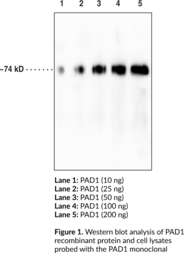
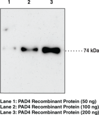
Protein arginine deiminases (PADs) are guanidine-modifying enzymes belonging to the amidinotransferase superfamily and are designated PAD1-4, and PAD6. PADs are calcium-dependent enzymes that catalyze the post-translational modification of target proteins by converting arginine to citrulline.{19489, 18141} The excess deimination of target proteins can result in the production of anti-citrullinated protein antibodies (ACPAs) which can be indicators of a number of disease states.{31883} The various PADs exhibit tissue specific expression and different subcellular localization.{19488} PAD1 is expressed in uterus and throughout the epidermis. PAD1 and PAD3 are speculated to mediate deamination of epidermal filaggrin (filament aggregation protein) and keratins, proteins involved in maintaining skin hydration.{19490} The predicted size for PAD1 is 74.7 kD and Cayman’s PAD1 monoclonal antibody (clone 6B4) detects a band at ~74 kD by Western blot.
Brand:CaymanSKU:22997 - 100 µgAvailable on backorder
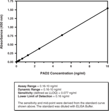
Peptidylarginine deiminases (PADs) are a family of five enzymes that catalyze the conversion of arginine to citrulline in peptides and proteins. PAD2 is the most widely expressed and conserved member across mammalian species, implying it is the ancestral homologue of the PADs. Cayman’s PAD2 (human) ELISA Kit is a sandwich assay that can be used to measure PAD2 in tissue culture medium, cell lysates, plasma, and serum.
Brand:CaymanSKU:501450 - 96 wellsAvailable on backorder
Protein arginine deiminase 2 (PAD2) is a guanidino-modifying enzyme that catalyzes the conversion of specific arginine residues to citrulline in a calcium-dependent manner. This enzyme has been implicated in several diseases, including rheumatoid arthritis (RA), retinal degeneration, and certain cancers. PAD2 has been shown to modify vimentin, fibrinogen, and β/γ-actin. Extracellular levels of PAD2 are increased in the lungs of smokers, providing a link between smoking as a risk factor for RA and anti-citrullinated protein antibodies among RA patients. Cayman’s PAD2 Inhibitor Screening Assay Kit (AMC) provides a convenient method for screening human PAD2 inhibitors.
Brand:CaymanSKU:701390 - 96 wellsAvailable on backorder
Protein Arginine Deiminase 2 (PAD2) is a guanidino-modifying enzyme that catalyzes the conversion of specific arginine residues to citrulline in a calcium-dependent manner. This enzyme has been implicated in several diseases, including rheumatoid arthritis (RA), retinal degeneration, and certain cancers. PAD2 has been shown to modify vimentin, fibrinogen and β/γ-actin. Extracellular levels of PAD2 are increased in the lungs of smokers, providing a link between smoking as a risk factor for RA and anti-citrullinated protein antibodies among RA patients. Cayman’s PAD2 Inhibitor Screening Assay (Ammonia) provides a convenient method for screening human PAD2 inhibitors.
Brand:CaymanSKU:701400 - 96 wellsAvailable on backorder
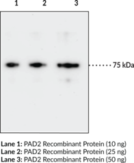
Protein Arginine Deiminases (PADs) are guanidino-modifying enzymes belonging to the amidinotransferase superfamily and are designated PAD1-4 and PAD6. PAD enzymes catalyze the conversion of specific arginine residues to citrulline in a calcium-dependent manner. All enzymes are cytosolic except for PAD4 which is localized in the nucleus.{18141} PAD2 is the most widely expressed member and also the most conserved across mammalian species, implying it is the ancestral homologue of the PADs.{19489} Overexpression of PAD2 results in myelin loss in a transgenic model, potentially linking PAD2 activity to multiple sclerosis.{30727} It has also been shown to modify vimentin and β/γ-actin, potentially aggravating the autoantigen response in rheumatoid arthritis.{30728,30130} PAD2 may also play a role in transcriptional regulation, as it has been shown capable of citrullinating histones, particularly H3 during mammalian reproductive cycles, when it is transcriptionally activated in the nucleus.{30729} The predicted molecular weight of PAD2 is 75.6 kDa and Cayman’s PAD2 Monoclonal Antibody (Clone 9F7) detects a band at 75 kDa by western blot.
Brand:CaymanSKU:19822 - 100 µgAvailable on backorder
Cayman’s PAD3 Inhibitor Screening Assay Kit (Ammonia) provides a convenient method for screening human PAD3 inhibitors. PAD3 deiminates N-α-benzoyl-L-arginine ethyl ester (BAEE), a non-natural substrate with similar kinetic properties to the natural substrates, producing ammonia.{18133} Ammonia reacts with a detector resulting in a fluorescent product. Fluorescence is then analyzed with an excitation wavelength of 405-415 nm and an emission wavelength of 470-480 nm.
Brand:CaymanSKU:701470 - 96 wellsAvailable on backorder
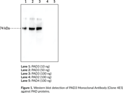
Protein arginine deiminase 3 (PAD3; Item No. 10786) is a homodimeric guanidine-modifying enzyme belonging to the amidinotransferase superfamily.{34555} It is a calcium-dependent enzyme that catalyzes the post-translational modification of target proteins by converting arginine to citrulline. Substrates of PAD3 include filaggrin, trichohyalin, apoptosis-inducing factor (AIF), vimentin, and other proteins.{34556} PAD3 is expressed in the epidermis, hair follicles, and keratinocytes. It has a role in shaping and mechanical strengthening of hair, and mutations in PAD3 lead to uncombable hair syndrome.{36391} PAD3 is also expressed in mammary glands and citrullinates proteins during lactation.{34764} Cayman’s PAD3 Monoclonal Antibody (Clone 4E5) can be used for Western blot and ELISA. The predicted size for PAD3 is 76.4 kDa and Cayman’s PAD3 Monoclonal Antibody (Clone 4E5) recognizes PAD3 at ~74 kDa from human samples.
Brand:CaymanSKU:24377 - 100 µgAvailable on backorder
Brand:CaymanSKU:700562 - 60 µlAvailable on backorder
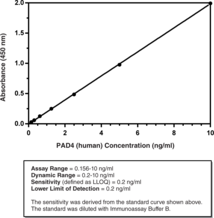
Peptidyl arginine deiminases (PADs) are a family of five enzymes that catalyzes the conversion of arginine to citrulline in peptides and proteins.{31654} PAD4 is in the nucleus of neutrophils and monocytes, and other hematopoietic cells. Cayman’s PAD4 (human) ELISA Kit is a sandwich assay that can be used to measure PAD4 in tissue culture medium, cell lysates, plasma, and serum.
Brand:CaymanSKU:501460 - 96 WellAvailable on backorder
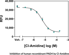
Protein arginine deiminase 4 (PAD4) is a guanidino-modifying enzyme that functions as a transcriptional coregulator catalyzing the conversion of specific arginine residues to citrulline. Substrates for PAD4 include histones H2A, H3, and H4. PAD4 autocitrullinates itself at several sites, inhibiting its enzymatic activity. PAD4 activity is increased in rheumatoid arthritis, producing an abundance of citrulline-containing proteins that generate an immune response resulting in production of autoantibodies that ultimately attack the host tissues. PAD4 has also been implicated in several other diseases including multiple sclerosis, Alzheimer’s disease, glaucoma, and cancer. Cayman’s PAD4 Inhibitor Screening Assay (AMC) provides a convenient, fluorescence-based method for screening human PAD4 inhibitors.
Brand:CaymanSKU:701320 - 96 wellsAvailable on backorder
Protein Arginine Deiminase 4 (PAD4) is a guanidino-modifying enzyme that functions as a transcriptional coregulator catalyzing the conversion of specific arginine residues to citrulline. Substrates for PAD4 include histones H2A, H3, and H4. PAD4 autocitrullinates itself at several sites, inhibiting its enzymatic activity. PAD4 activity is increased in rheumatoid arthritis, producing an abundance of citrulline-containing proteins that generate an immune response resulting in production of autoantibodies that ultimately attack the host tissues. PAD4 has also been implicated in several other diseases including multiple sclerosis, Alzheimer’s disease, glaucoma, and cancer. Cayman’s PAD4 Inhibitor Screening Assay (Ammonia) provides a convenient, fluorescence-based method for screening human PAD4 inhibitors.
Brand:CaymanSKU:700560 - 96 wellsAvailable on backorder
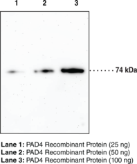
Protein arginine deiminases (PADs) are guanidino-modifying enzymes belonging to the amidinotransferase superfamily and are designated PAD1-4 and PAD6. All enzymes are cytosolic except for PAD4 which is localized in the nucleus.{18141} PAD4 is a homodimer that functions as a transcriptional coregulator to catalyze the conversion of specific arginine residues to citrulline in a calcium-dependent manner. PAD4 substrates include histones H2A, H3, and H4, whose post-translational modifications play a large role in gene regulation.{18134} Benzoylated arginine substrates like N-α-benzoyl-L-arginine ethyl ester have proven to be useful tools for characterization of PAD4, having similar kinetic properties to the natural substrates.{18133} PAD4 itself can undergo autocitrullination at several sites, which inhibits its enzymatic activity and may play an important role in regulating citrullination in cells.{18135} PAD4 activity is increased in rheumatoid arthritis, producing an abundance of citrulline-containing proteins that can be recognized by autoantibodies, causing the immune system to attack its own tissues.{18137} PAD4 has also been implicated in several other diseases including multiple sclerosis, Alzheimer’s, glaucoma, and cancer.{18134} The predicted size of PAD4 is 74.1 kDa and Cayman’s PAD4 Monoclonal Antibody (Clone 11F9) detects a band at 74 kDa by western blot.
Brand:CaymanSKU:19671 - 100 µgAvailable on backorder
Protein arginine deiminases (PADs) are guanidino-modifying enzymes belonging to the amidinotransferase superfamily and are designated PAD1-4 and PAD6. All enzymes are cytosolic except for PAD4 which is localized in the nucleus.{18141} PAD4 is a homodimer that functions as a transcriptional coregulator to catalyze the conversion of specific arginine residues to citrulline in a calcium-dependent manner. PAD4 substrates include histones H2A, H3, and H4, whose post-translational modifications play a large role in gene regulation.{18134} Benzoylated arginine substrates like N-α-benzoyl-L-arginine ethyl ester have proven to be useful tools for characterization of PAD4, having similar kinetic properties to the natural substrates.{18133} PAD4 itself can undergo autocitrullination at several sites, which inhibits its enzymatic activity and may play an important role in regulating citrullination in cells.{18135} PAD4 activity is increased in rheumatoid arthritis, producing an abundance of citrulline-containing proteins that can be recognized by autoantibodies, causing the immune system to attack its own tissues.{18137} PAD4 has also been implicated in several other diseases including multiple sclerosis, Alzheimer’s, glaucoma, and cancer.{18134} The predicted size of PAD4 is 74.1 kDa and Cayman’s PAD4 Monoclonal Antibody (Clone 6D8) detects a band at 74 kDa by western blot.
Brand:CaymanSKU:19669 - 100 µgAvailable on backorder
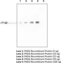
Protein arginine deiminase 6 (PAD6) is a homodimeric guanidine-modifying enzyme belonging to the amidinotransferase superfamily.{34555} It is a calcium-dependent enzyme that catalyzes the post-translational modification of target proteins by converting arginine to citrulline. PAD6 is expressed in mammalian oocytes, sperm cells, and early embryos.{39678} In mammalian oocytes and early embryo cytoplasm, its expression is localized to cytoskeletal sheets, dynamic structures containing various keratins, which are major targets for citrullination. PAD6-/- oocytes exhibit reduced microtubule acetylation and defective organelle positioning and redistribution, suggesting a role for PAD6 in regulating microtubule-mediated organelle movement and positioning.{39677} PAD6-/- female, but not male, mice are infertile due to a reduction of de novo protein synthesis, cytoskeletal sheet formation, and ribosomal RNA which induces arrest of zygote development at the two-cell stage.{39678,39679} PAD6 is regulated by newborn ovary homeobox (Nobox), as its promoter contains a Nobox DNA-binding element (NBE) and expression and activity of PAD6 are decreased in Nobox-/- mouse ovaries.{39676} In human females, homozygous nonsense mutations and compound-heterozygous mutations in PAD6 induce early embryonic arrest following in vitro fertilization (IVF) or intracytoplasmic sperm injection (ICSI).{39680} Cayman’s PAD6 monoclonal antibody (Clone 4B7) can be used for Western blot and ELISA applications. The antibody recognizes PAD6 at ~77 kDa from human samples.
Brand:CaymanSKU:25965 - 100 µgAvailable on backorder
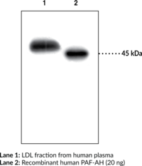
PAF Acetylhydrolase (PAF-AH) catalyzes the hydrolysis of the acetyl group at the sn-2 position of PAF to yield lyso-PAF and acetate, thereby inactivating PAF. Two main types of PAF-AH have been characterized, namely secreted (i.e., plasma) and intracellular enzymes, which are encoded on individual genes and share only 41% homology at the amino acid level.{4758,2618,6501} Plasma PAF-AH is a 45.4 kDa enzyme which is associated with the LDL fraction of plasma.{2618,2626}
Brand:CaymanSKU:160603 - 500 µlAvailable on backorder
Brand:CaymanSKU:760910 - 1 eaAvailable on backorder
Brand:CaymanSKU:760911 - 1 eaAvailable on backorder
Platelet-activating factor (PAF) is a biologically active phospholipid synthesized by a variety of cells upon stimulation. PAF is converted to the biologically inactive lyso-PAF by the enzyme PAF acetylhydrolase (PAF-AH). Plasma PAF-AH is highly selective for phospholipids with very short acyl groups at the sn-2 position and is associated with lipoproteins.{2626} The Cayman Chemical PAF acetylhydrolase assay kit provides an accurate and convenient method for measurement of PAF-AH activity. The assay uses 2-thio PAF which serves as a substrate for PAF-AH.{3787} Upon hydrolysis of the acetyl thioester bond at the sn-2 position by PAF-AH, free thiols are detected using 5,5′-dithio-bis(2-nitrobenzoic acid) (DTNB; Ellman’s reagent). The dynamic range of the assay is only limited by the accuracy of the absorbance measurement. Most plate readers are linear to an absorbance of 1.2. The detection range of the assay is from 0.02 to 0.2 µmol/min/ml of PAF-AH activity which is equivalent to an absorbance increase of 0.01 to 0.1 per minute. Each kit contains assay buffer, DTNB, 2-thio PAF (substrate), human PAF-AH standard, a 96 well plate, and complete instructions.
Brand:CaymanSKU:760901 - 96 wellsAvailable on backorder
Platelet-activating factor (PAF) is a biologically active phospholipid synthesized by a variety of stimulated cells. The biological effects of PAF include activation of platelets, polymorphonuclear leukocytes, monocytes, and macrophages. PAF also increases vascular permeability, decreases cardiac output, induces hypotension, and stimulates uterine contraction.{939} PAF has been implicated in pathological processes, such as inflammation and allergy.{11753} PAF is converted to the biologically inactive lyso-PAF by the enzyme PAF acetylhydrolase (PAF-AH). PAF-AHs are located intra- and extra-cellularly (e.g., cytosolic and plasma). Plasma PAF-AH is highly selective for phospholipids with very short acyl groups at the sn-2 position and is associated with lipoproteins.{2626} Recently, plasma PAF-AH has been linked to atherosclerosis and may be a positive risk factor for coronary heart disease in humans.{11455} The Cayman Chemical PAF Acetylhydrolase Inhibitor Screening Assay uses 2-thio PAF as a substrate for PAF-AH.{3787} Upon hydrolysis of the acetyl thioester bond at the sn-2 position by PAF-AH, free thiols are detected using 5,5′-dithio-bis-(2-nitrobenzoic acid) also known as DTNB or Ellman’s reagent. Cayman’s PAF-AH Inhibitor Screening assay includes human plasma PAF-AH and is a time saving tool for screening vast numbers of inhibitors.
Brand:CaymanSKU:10004380 - 96 wellsAvailable on backorder
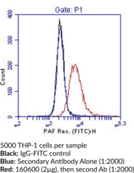
PAF is a potent phospholipid mediator which exerts diverse biological actions by interaction with a G protein-coupled PAF receptor. The PAF receptor has been cloned from a number of species including human, rat, and guinea pig and is characterized as a 7-transmembrane receptor which induces phosphoinositol turnover through G-protein coupling.{5109,5148,5150,5145,5149} Northern blot analysis reveals that the receptor is expressed in leukocytes, placenta, lung, spleen, small intestine, kidney, liver, and brain.{5150,5145} In leukocyte cell populations the receptor is found on platelets, monocytes, neutrophils, and B-cells, whereas resting T-cells and natural killer cell lines do not express the PAF receptor.{4226} Human monocytes treated with INF-γ have a 2-6 fold increase in PAF receptor expression compared to untreated cells.{4225}
Brand:CaymanSKU:160600 - 1 eaAvailable on backorder
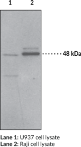
PAF is a potent phospholipid mediator which exerts diverse biological actions by interaction with a G protein-coupled PAF receptor. The PAF receptor has been cloned from a number of species including human, rat, and guinea pig and is characterized as a 7-transmembrane receptor which induces phosphoinositol turnover through G-protein coupling.{5109,5148,5150,5145,3513} Northern blot analysis reveals that the receptor is expressed in leukocytes, placenta, lung, spleen, small intestine, kidney, liver, and brain.{5150,5145} In leukocyte cell populations the receptor is found on platelets, monocytes, neutrophils, and B-cells, whereas resting T-cells and natural killer cell lines do not express the PAF receptor.{4226} Human monocytes treated with INF-γ have a 2-6 fold increase in PAF receptor expression compared to untreated cells.{4225} PAF receptor is detected on immunoblot at 48 kDa.{12339}
Brand:CaymanSKU:160602 - 1 eaAvailable on backorder
Brand:CaymanSKU:10004800 - 1 eaAvailable on backorder
Brand:CaymanSKU:10004801 - 1 eaAvailable on backorder
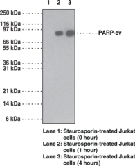
The poly(ADP-ribose) polymerase (PARP) is involved in cell recovery from DNA damage, such as methylation of N3-adenine, which activates the base excision repair process. PARP is a 116 kDa nuclear chromatin-associated enzyme that is cleaved during apoptosis by caspase-3 into a 24 kDa fragment containing the DNA binding domain and an 89 kDa fragment containing the catalytic and automodification domains. The 24 kDa fragment irreversibly binds to DNA and may contribute to the irreversibility of apoptosis by blocking the access of DNA repair enzymes to DNA strand breaks.
Brand:CaymanSKU:13557 - 1 eaAvailable on backorder
Paxillin is a focal adhesion adapter protein that facilitates the assembly of multiprotein complexes that mediate signal transduction between the extracellular matrix and actin cytoskeleton.{59175,59176,59177} It contains four LIM domains in the C-terminal region that target paxillin to focal adhesions and an N-terminal region with five LD motifs that serve as docking sites for a variety of proteins, including vinculin, focal adhesion kinase (FAK), and Src, that facilitate intracellular signal transduction.{59177} Alternative splicing of PXN generates three isoforms, α, β, and γ, that contain a variable N-terminus, as well as a fourth isoform, δ, that lacks the N-terminal region.{59177} Paxillin is expressed in most tissues and predominately localizes to focal adhesions on the cell membrane.{59176,59177} Upon cytokine or growth factor stimulation or cell adhesion, paxillin is phosphorylated by a variety of kinases, including FAK and ERK, providing a scaffold for signaling proteins that are involved in the formation and regulation of focal adhesions, which mediate cell migration.{59177} It is also expressed in the cytoplasm and nucleus where it regulates gene transcription. Tumor paxillin levels are increased in patients with glioblastoma multiforme (GBM) and associated with poor survival. Cayman’s Paxillinα/β/γ (N-Term) Rabbit Monoclonal Antibody can be used for immunocytochemistry (ICC) and Western blot (WB) applications.
Brand:CaymanSKU:32209 - 100 µlAvailable on backorder
PBS Assay Buffer (10X) has been tested and formulated to work exclusively with Cayman’s Protein Aggregation Fluorometric Assay Kit (Item No. 701760). Please visit Protein Aggregation Fluorometric Assay Kit (Item No. 701760) for the kit protocol, procedures, and product handling.
Brand:CaymanSKU:700031 - 5 mlAvailable on backorder
The proliferating cell nuclear antigen (PCNA) becomes detectable in the G1 phase of the cell cycle and reaches a maximum level of expression during mitosis. PCNA is highly conserved across species and is a critical part of the DNA polymerase δ holoenzyme.{12084,12085,12086} The level of PCNA expression depends on the proliferative potential of the cell examined. Caymanl’s PCNA Monoclonal Antibody can be used for analysis of PCNA by Western blot, flow cytometry, immunocytochemistry, and immunohistochemistry (frozen and paraffin-embedded tissue) on samples from multiple species. This monoclonal antibody stains proliferating cells in a wide range of normal tissues, as well as in tissues undergoing pathological processes and after mitogenic and allogeneic stimulations.
Brand:CaymanSKU:10004805 - 1 eaAvailable on backorder
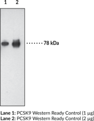
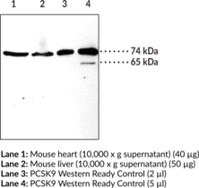
Proprotein convertase subtilisin kexin 9 (PCSK9) is a member of the subtilisin serine protease family with an important role in lipoprotein metabolism.{13178} Gain-of-function mutations in the PCSK9 gene are associated with autosomal dominant hypercholesterolemia which is characterized by an increase in low-density lipoprotein (LDL) cholesterol levels.{13158} PCSK9 overexpression in wild-type mice doubles the plasma total cholesterol, possibly through accelerated LDL receptor degredation.{13178,13176} PCSK9 mRNA is detected in various tissues such as liver, kidney, lung, spleen, jejunum, ileum, colon, and muscle with the highest expression in the liver.{13136} Human PCSK9 precursor is 692 amino acids in length with an estimated molecular weight of 74 kDa. This proprotein is self-cleaved to form a mature protein at around 63 kDa in the Golgi body.{13177}
Brand:CaymanSKU:10218 - 1 eaAvailable on backorder
PCSK9 is a member of the subtilisin serine protease family with an important role in low-density lipoprotein (LDL) metabolism.{13178} Mutation in the PCSK9 gene is associated with autosomal dominant hypercholesterolemia which is characterized by an increase in LDL cholesterol levels.{13158} PCSK9 overexpression in wild-type mice doubles the plasma total cholesterol, possibly through acceleration of the degradation of the LDL receptor.{13176,13178} PCSK9 mRNA is detected in various tissues such as liver, kidney, lung, spleen, jejunum, ileum, colon, and muscles with the highest expression in the liver.{13136} Human PCSK9 precursor is 692 amino acids in length with an estimated molecular weight of 74 kDa. This proprotein is self-cleaved to form a mature protein of 63 kDa in the Golgi.{13177}
Brand:CaymanSKU:10240 - 1 eaAvailable on backorder
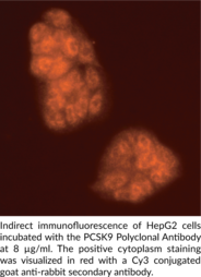
Proprotein convertase subtilisin kexin 9 (PCSK9) is a member of the subtilisin serine protease family with an important role in lipoprotein metabolism.{13178} Mutation in the PCSK9 gene is associated with autosomal dominant hypercholesterolemia which is characterized by an increase in low density lipoprotein (LDL) cholesterol levels.{13158} PCSK9 overexpression in wild-type mice doubles the plasma total cholesterol, possibly through acceleration of the degradation of the LDL receptor.{13176,13178} PCSK9 mRNA is detected in various tissues such as liver, kidney, lung, spleen, jejunum, ileum, colon, and muscle with the highest expression in the liver.{13136} Human PCSK9 precursor is 692 amino acids in length with an estimated molecular weight of 74 kDa. This proprotein is self-cleaved to form a mature protein at around 63 kDa in the golgi.{13177} Cayman’s PCSK9 polyclonal antibody detects mainly the mature form of the protein ranging from 62-66 kDa in tissues and cells such as liver, kidney, and colon cancer cells.
Brand:CaymanSKU:10007185 - 500 μlAvailable on backorder
Proprotein convertase subtilisin kexin 9 (PCSK9) is a member of the subtilisin serine protease family with an important role in lipoprotein metabolism.{13178} Mutation in the PCSK9 gene is associated with autosomal dominant hypercholesterolemia which is characterized by an increase in low density lipoprotein (LDL) cholesterol levels.{13158} PCSK9 overexpression in wild-type mice doubles the plasma total cholesterol, possibly through acceleration of the degradation of the LDL receptor.{13176,13178} PCSK9 mRNA is detected in various tissues such as liver, kidney, lung, spleen, jejunum, ileum, colon, and muscle with the highest expression in the liver.{13136} Human PCSK9 precursor is 692 amino acid in length with an estimated molecular weight of 74 kDa. This proprotein is self-cleaved to form a mature protein at around 63 kDa in the Golgi.{13177}
Brand:CaymanSKU:10008811 - 500 µlAvailable on backorder
The three dimensional structure of many extracellular proteins is stabilized by the formation of disulphide bonds. Studies suggest that a microsomal enzyme known as protein disulphide isomerase (PDI) is involved in disulphide-bond formation via its oxidase activity and isomerization via its isomerase activity, as well as the reduction of disulphite bonds in proteins.{16242} Studies suggest BiP and PDI work together sequentially to increase oxidation of these proteins.{16273,16274} PDI has also been found to function as a chaperone to prevent the aggregation of unfolded substrates, and serves as a subunit of prolyl 4-hydroxylase and microsomal triglyceride transferase.{16275,16276} PDI is an abundant 55 kDa protein located primarily in the ER, however studies have also proved its presence in the cytosol.{16272} PDI has the ability to reside in the ER permanently due to the highly conserved KDEL sequence at its carboxy-terminus.{16277} It uses carboxy-terminal KDEL as a retention signal, and this appears to be sufficient to reduce the secretion of proteins from the ER. This retention is reported to be mediated by a KDEL receptor.{16278}
Brand:CaymanSKU:13025 - 100 µlAvailable on backorder
The three dimensional structure of many extracellular proteins is stabilized by the formation of disulphide bonds. Studies suggest that a microsomal enzyme known as protein disulphide isomerase (PDI) is involved in disulphide-bond formation via its oxidase activity and isomerization via its isomerase activity, as well as the reduction of disulphite bonds in proteins.{16242} Studies suggest BiP and PDI work together sequentially to increase oxidation of these proteins.{16273,16274} PDI has also been found to function as a chaperone to prevent the aggregation of unfolded substrates, and serves as a subunit of prolyl 4-hydroxylase and microsomal triglyceride transferase.{16275,16276} PDI is an abundant 55 kDa protein located primarily in the ER, however studies have also proved its presence in the cytosol.{16272} PDI has the ability to reside in the ER permanently due to the highly conserved KDEL sequence at its carboxy-terminus.{16277} It uses carboxy-terminal KDEL as a retention signal, and this appears to be sufficient to reduce the secretion of proteins from the ER. This retention is reported to be mediated by a KDEL receptor.{16278}
Brand:CaymanSKU:13025 - 25 µlAvailable on backorder
Phosphoenolpyruvate carboxykinase (PEPCK) is a hormonally regulated enzyme responsible for the first committed step in gluconeogenesis, catalyzing the conversion of oxaloacetate to phosphoenolpyruvate.{13231,13234} PEPCK mRNA is most abundant in the liver, kidney cortex, and white adipose tissue but is also found in brown fat, the mammary gland during lactation, and the small intestine.{13234} Both non-insulin-dependent diabetes mellitus and streptozotocin-induced diabetes result in elevated PEPCK activity, protein, and mRNA.{13231,13237} Two isozymes of PEPCK have been identified, cytosolic and mitochondrial.{13240} Cayman’s PEPCK polyclonal antibody was generated against murine cytosolic PEPCK and detects the protein at 63 kDa by western blotting in rat and murine samples.
Brand:CaymanSKU:10004943 - 1 eaAvailable on backorder
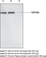
Cayman’s PHLPP1 Polyclonal Antibody detects the carboxy-terminal amino acids within the PDZ-binding domain of both the PHLPP1 α and β isoforms. PHLPP2 shares partial identity with the domains of PHLPP1 however it has a unique PDZ-binding domain sequence. Protein kinase B/Akt is critical in regulating cell growth and death. Activation of a serine residue (Ser473) in a C-terminal hydrophobic motif of Akt is known to be linked to some of the most common human cancers. PHLPP1 and PHLPP2 dephosphorylate the hydrophobic motif of Akt and thus reduce Akt activity, resulting in a increase in the number of apoptotic cells.{13717} PHLPP1 levels are markedly reduced in several colon cancer and glioblastoma cell lines that have elevated Akt phosphorylation. Injection of a human glioblastoma cell line overexpressing PHLPP1 into mice reduced tumor size by nearly 70% compared with control animals. mRNA of PHLPP1 is ubiquitously expressed, with highest levels found in brain.{13717} Cayman’s PHLPP Polyclonal Antibody detects the enzyme at around 150 kDa (α isoform) and 200 kDa (β isoform) by western blot from various cell lines (THP-1) and tissues such as brain.
Brand:CaymanSKU:10007191 - 1 eaAvailable on backorder
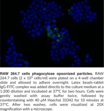
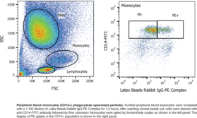
Cayman’s Phagocytosis Assay Kit (IgG FITC) employs latex beads coated with fluorescently-labeled rabbit IgG as a probe for the measurement of the phagocytic process in vitro. The engulfed fluorescent beads can be detected using a fluorescence microscope, allowing kinetic studies of phagocytosis at the single-cell level. In addition, the flow cytometric readout provides the advantage of visualizing perturbations in phagocytosis on the population level and, when combined with antibody staining, of specific cell types within complex populations. This kit provides enough Latex Beads-Rabbit IgG-FITC Complex for up to 750 samples.
Brand:CaymanSKU:500290 - 1 eaAvailable on backorder
Cayman’s Phagocytosis Assay Kit (IgG PE) employs latex beads coated with fluorescently-labeled rabbit IgG as a probe for the measurement of the phagocytic process in vitro. The engulfed fluorescent beads can be detected using a fluorescence microscope, allowing kinetic studies of phagocytosis at the single-cell level. In addition, the flow cytometric readout provides the advantage of visualizing perturbations in phagocytosis on the population level and, when combined with antibody staining, of specific cell types within complex populations. This kit provides enough Latex Beads-Rabbit IgG-PE Complex for up to 750 samples.
Brand:CaymanSKU:600540 - 1 eaAvailable on backorder
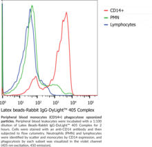
Cayman’s Phagocytosis Assay Kit (IgG-DyLight™ 405) employs latex beads coated with fluorescently-labeled rabbit IgG as a probe for the measurement of the phagocytic process in vitro. The engulfed fluorescent beads can be detected using a fluorescence microscope, allowing kinetic studies of phagocytosis at the single-cell level. In addition, the flow cytometric readout provides the advantage of visualizing perturbations in phagocytosis on the population level and, when combined with antibody staining, of specific cell types within complex populations. This kit provides enough Latex Beads-Rabbit IgG-DyLight™ 405 Complex for up to 500 samples.
Brand:CaymanSKU:601480 - 1 eaAvailable on backorder
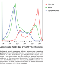
Cayman’s Phagocytosis Assay Kit (IgG-DyLight™ 633) employs latex beads coated with fluorescently-labeled rabbit IgG as a probe for the measurement of the phagocytic process in vitro. The engulfed fluorescent beads can be detected using a fluorescence microscope, allowing kinetic studies of phagocytosis at the single-cell level. In addition, the flow cytometric readout provides the advantage of visualizing perturbations in phagocytosis on the population level and, when combined with antibody staining, of specific cell types within complex populations. This kit provides enough Latex Beads-Rabbit IgG-DyLight™ 633 Complex for up to 500 samples.
Brand:CaymanSKU:601490 - 1 eaAvailable on backorder
Cayman’s phosphatidylcholine (PC) assay kit provides a specific, sensitive, and convenient method for quantifying PC in plasma or serum. In this assay, PC-Specific PLD is first used to hydrolyze PC to choline and phosphatidic acid. The newly formed choline is then used to generate hydrogen peroxide in a reaction catalyzed by choline oxidase. Finally, with peroxidase as a catalyst, hydrogen peroxide reacts with DAOS and 4-aminoantipyrine to generate a blue dye with an optimal absorption at 595 nm.{14806}
Brand:CaymanSKU:10009926 - 96 wellsAvailable on backorder
Phospholipase C β2 (PLCβ2) is an enzyme that catalyzes the hydrolysis of phosphatidylinositol 4,5-bisphosphate to the secondary messengers inositol 1,4,5-triphosphate (IP3) and diacylglycerol (DAG).{59155,59156} It is approximately 135 kDa and is composed of a pleckstrin homology domain, four EF-hand motifs, a catalytic domain, and a C2 domain that are common to all PLCs, as well as a 400-amino acid C-terminal region that is required for PLCβ2 activation by the G-protein subunits Gαq or Gα11.{59157} PLCβ2 is also activated by Gβγ subunits. Upon activation by a G-protein subunit, PLCβ2 initiates intracellular signal transduction of extracellular signals using calcium as a cofactor. PLCβ2 colocalizes with T1R and T2R taste receptors and Plcb2-/- mice exhibit selective and complete loss of sweet, amino acid, and bitter tastes.{59158} Plcb2-/- mice also exhibit increased coxsackievirus A16-induced pro-inflammatory cytokine production and decreased survival compared with wild-type mice.{59159} Cayman’s Phospholipase C β2 Polyclonal Antibody can be used for immunohistochemistry (IHC) and immunofluorescence (IF) applications.
Brand:CaymanSKU:29290 - 100 µlAvailable on backorder
Protein phosphorylation is an important post-translational modification that serves many key functions to regulate a protein’s activity, localization, and protein-protein interactions. Phosphorylation is catalyzed by various specific protein kinases, which involves removing a phosphate group from ATP and covalently attaching it to a recipient protein that acts as a substrate. Most kinases act on both serine and threonine; others act on tyrosine, and a number (dual specificity kinases) act on all three. Because phosphorylation can occur at multiple sites on any given protein, it can therefore change the function or localization of that protein at any time.{17293} Changing the function of these proteins has been linked to a number of diseases, including cancer, diabetes, heart disease, inflammation, and neurological disorders.{17295,17296,17297}
Brand:CaymanSKU:10221 - 100 µlAvailable on backorder
Phosphothreonine Monoclonal Antibody is a probe for immunochemical detection of phosphorylated threonine residues on proteins by immunoblotting, immunohistochemistry, or immunocytochemistry. The phosphorylation of threonine residues by serine/threonine kinases serves a variety of purposes, including altering activity, stability, and interaction with other biomolecules. Phosphorylation may be persistent or transient, with dephosphorylation mediated by phosphatases.
Brand:CaymanSKU:20716 - 100 µgAvailable on backorder
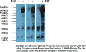
Protein phosphorylation is an important post-translational modification that serves many key functions to regulate a protein’s activity, localization, and protein-protein interactions. Phosphorylation is catalyzed by various specific protein kinases, which involves removing a phosphate group from ATP and covalently attaching it to a recipient protein that acts as a substrate. Most kinases act on both serine and threonine; others act on tyrosine, and a number (dual specificity kinases) act on all three. Because phosphorylation can occur at multiple sites on any given protein, it can therefore change the function or localization of that protein at any time.{17293} Changing the function of these proteins has been linked to a number of diseases, including cancer, diabetes, heart disease, inflammation, and neurological disorders.{17295,17296,17297}In particular, the phosphorylation of tyrosine is considered one of the key steps in signal transduction and regulation of enzymatic activity.{17301} Specific antibodies can detect phosphotyrosine and are therefore helpful for facilitating the identification of tyrosine kinase substrates in vivo.{17302}
Brand:CaymanSKU:10222 - 100 µlAvailable on backorder
Phosphotyrosine Monoclonal Antibody is a probe for immunochemical detection of phosphorylated tyrosine residues on proteins by immunoblotting. The phosphorylation of tyrosine residues by tyrosine kinases serves a variety of purposes, including altering activity, stability, and interaction with other biomolecules. Phosphorylation may be persistent or transient, with dephosphorylation mediated by phosphatases.
Brand:CaymanSKU:20717 - 100 µgAvailable on backorder
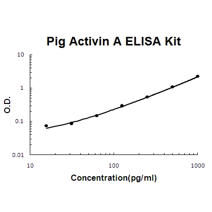
Sandwich High Sensitivity ELISA kit for Quantitative Detection of Pig porcine Activin A. 96wells/kit, with removable strips.
Brand:Boster BioSKU:EK0301-POAvailable on backorder
Sandwich High Sensitivity ELISA kit for Quantitative Detection of Pig porcine BDNF. 96wells/kit, with removable strips.
Brand:Boster BioSKU:EK0307-POAvailable on backorder
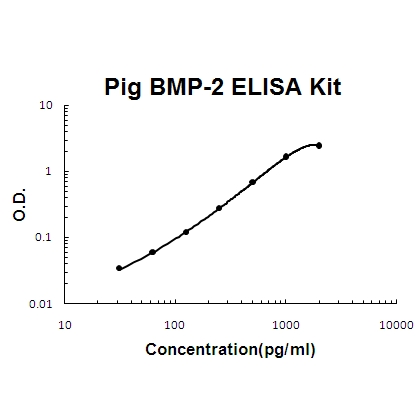
Sandwich High Sensitivity ELISA kit for Quantitative Detection of Pig porcine BMP-2. 96wells/kit, with removable strips.
Brand:Boster BioSKU:EK0311-POAvailable on backorder
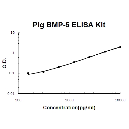
Sandwich High Sensitivity ELISA kit for Quantitative Detection of Pig porcine BMP-5. 96wells/kit, with removable strips.
Brand:Boster BioSKU:EK0310-POAvailable on backorder
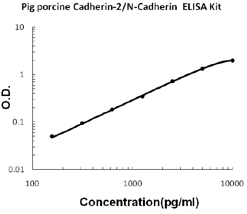
Sandwich High Sensitivity ELISA kit for Quantitative Detection of Pig porcine Cadherin-2/N-Cadherin. 96wells/kit, with removable strips.
Brand:Boster BioSKU:EK0669-POAvailable on backorder
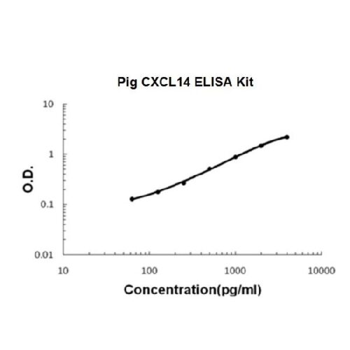
Sandwich High Sensitivity ELISA kit for Quantitative Detection of Pig porcine CXCL14. 96wells/kit, with removable strips.
Brand:Boster BioSKU:EK1285-POAvailable on backorder
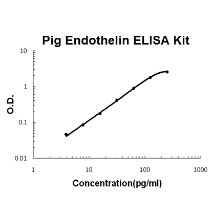
Sandwich High Sensitivity ELISA kit for Quantitative Detection of Pig porcine Endothelin. 96wells/kit, with removable strips.
Brand:Boster BioSKU:EK0945-POAvailable on backorder
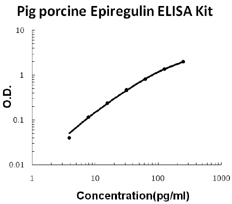
Sandwich High Sensitivity ELISA kit for Quantitative Detection of Pig porcine Epiregulin. 96wells/kit, with removable strips.
Brand:Boster BioSKU:EK1394-POAvailable on backorder
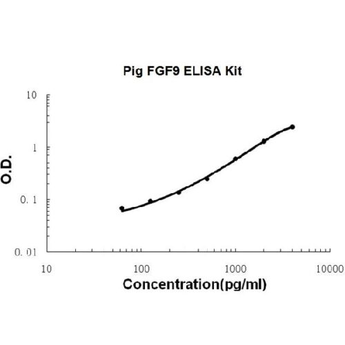
Sandwich High Sensitivity ELISA kit for Quantitative Detection of Pig porcine FGF9. 96wells/kit, with removable strips.
Brand:Boster BioSKU:EK0348-POAvailable on backorder
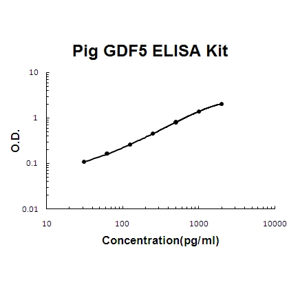
Sandwich High Sensitivity ELISA kit for Quantitative Detection of Pig porcine GDF5. 96wells/kit, with removable strips.
Brand:Boster BioSKU:EK1504-POAvailable on backorder
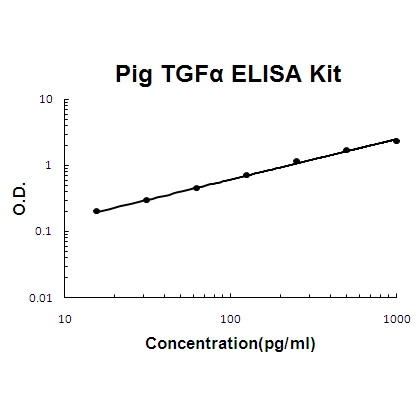
Sandwich High Sensitivity ELISA kit for Quantitative Detection of Pig porcine TGF alpha. 96wells/kit, with removable strips.
Brand:Boster BioSKU:EK0511-POAvailable on backorder
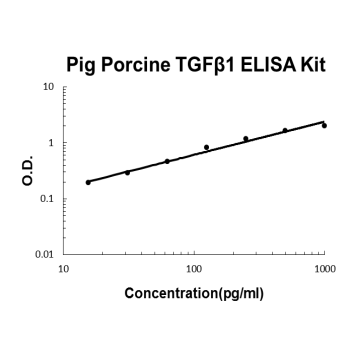
Sandwich High Sensitivity ELISA kit for Quantitative Detection of activated Pig porcine TGF beta 1. 96wells/kit, with removable strips.
Brand:Boster BioSKU:EK0513-POAvailable on backorder
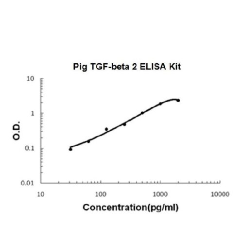
Sandwich High Sensitivity ELISA kit for Quantitative Detection of activated Pig porcine TGF-beta 2. 96wells/kit, with removable strips.
Brand:Boster BioSKU:EK0981-POAvailable on backorder
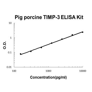
Sandwich High Sensitivity ELISA kit for Quantitative Detection of Pig porcine TIMP-3. 96wells/kit, with removable strips.
Brand:Boster BioSKU:EK0523-POAvailable on backorder
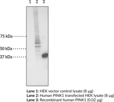
PTEN-induced putative kinase 1 (PINK1) is a serine/threonine protein kinase that has a role in mitochondrial function.{54198,54199} It is comprised of an N-terminal mitochondrial targeting sequence, a transmembrane domain, a serine/threonine kinase domain, and a C-terminal region.{54199} PINK1 is ubiquitously expressed primarily in the brain, skeletal muscle, and heart.{54200} It localizes to the mitochondria where it is either rapidly degraded or, under conditions of low mitochondrial membrane potential, accumulates on the outer mitochondrial membrane, where it recruits and activates the cytosolic E3 ubiquitin ligase Parkin, which targets the mitochondria for mitophagy.{54198,54200} Pink1 knockout in rats leads to an age-dependent loss of dopaminergic neurons in the substantia nigra, as well as deficits in motor function and mitochondrial respiration.{54201} In mice, Pink1 knockout does not induce a loss of dopaminergic neurons without concomitant overexpression of α-synuclein in the substantia nigra.{54202} Loss-of-function mutations in PINK1 are causally associated with autosomal recessive early-onset Parkinson’s disease.{54200,12321} Cayman’s PINK1 Monoclonal Antibody recognizes primarily the full length protein at about 66 kDa in human tissues. This antibody can be used for ELISA and Western blot applications.
Brand:CaymanSKU:29129 - 200 µgAvailable on backorder
PTEN-induced putative kinase 1 (PINK1) is a serine/threonine protein kinase that has a role in mitochondrial function.{54198,54199} It is comprised of an N-terminal mitochondrial targeting sequence, a transmembrane domain, a serine/threonine kinase domain, and a C-terminal region.{54199} PINK1 is ubiquitously expressed primarily in the brain, skeletal muscle, and heart.{54200} It localizes to the mitochondria where it is either rapidly degraded or, under conditions of low mitochondrial membrane potential, accumulates on the outer mitochondrial membrane, where it recruits and activates the cytosolic E3 ubiquitin ligase Parkin, which targets the mitochondria for mitophagy.{54200,54198} Pink1 knockout in rats leads to an age-dependent loss of dopaminergic neurons in the substantia nigra, as well as deficits in motor function and mitochondrial respiration.{54201} In mice, Pink1 knockout does not induce a loss of dopaminergic neurons without concomitant overexpression of α-synuclein in the substantia nigra.{54202} Loss-of-function mutations in PINK1 are causally associated with autosomal recessive early-onset Parkinson’s disease.{54200,12321} Cayman’s PINK1 Polyclonal Antibody can be used for immunohistochemistry (IHC) and Western blot (WB) applications. The antibody primarily recognizes full-length PINK1 at approximately 66 kDa from human, mouse, and rat samples but also detects a truncated form of the protein at approximately 33 kDa.
Brand:CaymanSKU:10006283 - 1 eaAvailable on backorder
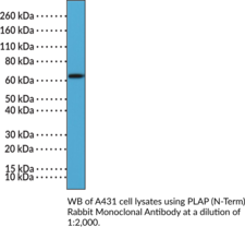
Placental alkaline phosphatase (PLAP) is the placenta-specific isoenzyme of AP encoded by the ALPP gene in humans.{59555,59556} It exists as a homodimer where each monomer is composed of an N-terminal α-helix and a crown domain that stabilize PLAP homodimers, a Zn2+- and Mg2+-containing active site with a catalytic serine residue, and a non-catalytic metal-binding site occupied by Ca2+.{59558,59557} PLAP is predominately expressed by syncytiotrophoblasts in the placenta and is tethered to the plasma membrane by a glycosylphosphatidylinositol (GPI) anchor.{59557,59558} It is also present at low levels in the cervix, ovaries, and type I pneumocytes, as well as in the serum.{59555,59558} Increased PLAP activity has been found in tumor tissue and cerebrospinal fluid (CSF) isolated from patients with intracranial germ cell tumors.{59559} Cayman’s PLAP (N-Term) Rabbit Monoclonal Antibody can be used for immunohistochemistry (IHC) and Western blot (WB) applications.
Brand:CaymanSKU:32259 - 100 µlAvailable on backorder
Brand:CaymanSKU:400145 - 1 eaAvailable on backorder
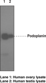
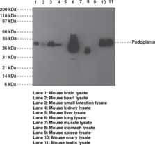
Podoplanin is a mucin type-1 glycoprotein of 40-43. Podoplanin is expressed in many tumors and many normal cells, especially lymphatic epithelial cells and follicular DCs. It appears to serve as a ligand for CLEC-2. Podoplanin expression is positively correlated with tumors expressing greater invasive and metastatic potential. Podoplanin is directly involved in cell migration, aids metastases formation and tumor cell invasion of tissue.{19761,19762,19763} It has also been determined that podoplanin expression is induced through Src activation of Cas.{19760} Recently there are indications of podoplanin presence on Th17 cells.{19759} Initial studies below indicate the presence of podoplanin on a small but distinct population of CD4+ cells from human PBMC.
Brand:CaymanSKU:11004 - 1 eaAvailable on backorder
Podoplanin is a 43 kD type 1 transmembrane glycoprotein found on many tissues. Podoplanin appears to be differentially expressed by lymphatic endothelial cells but not blood vessel endothelial cells.{19761} Podoplanin has also been found to be expressed on a wide variety of tumors. It appears to be involved in lymphangioigenesis and promoting cell migration.{19764} Recently, there are indications of podoplanin presence on Th17 cells.{19759} In a murine EAE model for autoimmune disease it has been shown that Th17 cells migrate quickly to the CNS at EAE onset. Injection of antibody to podoplanin in mice also immunized with myelin antigen to induce EAE results in elevated numbers of Th17 cells in the CNS. Thus as in effecting migration of tumor cells, podoplanin may also promote migration of committed Th17 cells to sites of inflammation in autoimmune diseases.
Brand:CaymanSKU:11005 - 1 eaAvailable on backorder
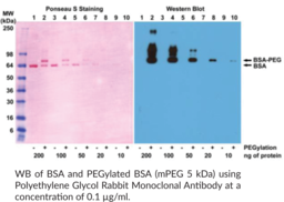
Polyethylene glycols (PEGs) are synthetic and hydrophilic polymers.{57302,57303} They are linear or branched and contain a reactive end group, such as acrylate, methacrylate, dibenzocyclooctynol, or vinyl sulfonate, for covalent attachment to macromolecules or linkers. The opposite end group of PEGs is commonly a methyl group (methoxy PEG), however, hydroxy, amino, butoxy, and tert-butoxy end groups have also been used.{57302} PEGs are non-toxic and are commonly used to prolong the in vivo circulation time of pharmaceutical agents.{57303} Free PEGs are non-immunogenic but become immunogenic when conjugated to a drug delivery nanosystem (DDS) or a macromolecule.{57302} Immunogenicity of PEGs varies based on polymer length and branching, end group composition, and chemical nature of the PEG acceptor structure. Cayman’s Polyethylene Glycol Rabbit Monoclonal Antibody can be used for ELISA, immunohistochemistry (IHC), and Western blot (WB) applications. The antibody recognizes PEGs containing a methoxy end group.
Brand:CaymanSKU:32180 - 100 µgAvailable on backorder
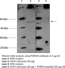
PORCN is a porcupine homolog protein that belongs to the evolutionary conserved porcupine gene family.{23738,23740} Genes of this family encode multi-pass endoplasmic reticulum proteins.{23740} Porcupine proteins play an important role in regulating Wnt signaling.{23738} Deficiency or mutation in X-linked PORCN causes focal dermal hypoplasia.{23739,23741}
Brand:CaymanSKU:14702 - 1 eaAvailable on backorder
Brand:CaymanSKU:700517 - 5 mlAvailable on backorder
Brand:CaymanSKU:32801 - 100 µlAvailable on backorder
